Download
1 / 27
270 likes | 409 Views
Abdomen 3. 2.6 Jejenum, ileum and colon. Albert van Schoor GNK 288 (SA4 Anatomy dissection). 2.6 Jejenum, ileum and colon. 2.6.1 Surface anatomy 2.6.2 Structure 2.6.3 Blood supply and lymph drainage 2.6.4 Embryology 2.6.5 Radiographic anatomy. 2.6.1 Surface anatomy.

E N D
Abdomen 3 2.6 Jejenum, ileum and colon Albert van Schoor GNK 288 (SA4 Anatomy dissection)
2.6 Jejenum, ileum and colon 2.6.1 Surface anatomy 2.6.2 Structure 2.6.3 Blood supply and lymph drainage 2.6.4 Embryology 2.6.5 Radiographic anatomy
2.6.1 Surface anatomy • Identify and describe in which region the jejunum and ileum are found • Review in which region the ascending, transverse and descending colon, caecum, appendix and sigmoid colon are found
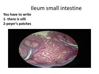
2.6.1 Surface anatomy Jejenum & ileum
2.6.1 Surface anatomy Colon
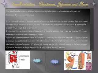
2.6.2 Structure • Identify and name the origin and termination of the jejunum and ileum • Identify the general and peritoneal relations of the jejunum and ileum • Compare and identify the blood supply, mucosal pattern, mesenterium and cross section of the proximal jejunum and distal ileum
2.6.2 Structure Proximal Jejenum Distal ileum
2.6.2 Structure • Compare and identify the differences between the small intestine and colon • Name and identify the duodenojejunal and ileocolic junctions and the root of the mesentery running between these points
Teniae coli Haustra Omental appendices / Appendices epiploica Semicircular folds Large diameter No teniae coli No Haustra No Omental appendices / Appendices epiploica Circular folds Small diameter 2.6.2 Structure DifferencesLarge & small intestines
2.6.2 Structure • Identify and briefly discuss the division of the large intestine into the caecum, appendix, ascending colon, transverse colon, descending colon and sigmoid colon, their structure and general and peritoneal relationships
2.6.2 Structure Cecum & appendix
2.6.2 Structure Ascending colon
2.6.2 Structure Transverse colon
2.6.2 Structure Descending colon
2.6.2 Structure Sigmoid colon
2.6.2 Structure • Briefly discuss and identify the anatomy of the appendix regarding its various positions, mesentery, blood supply and surface anatomy
2.6.2 Structure Appendix
2.6.3 Blood supply and lymph drainage • Schematically illustrate the arterial blood supply of the ileum, jejunum and colon • Name and identify the superior and inferior mesenteric vessels • Name and identify the arterial anastomoses between the foregut and midgut and midgut and hindgut
2.6.3 Blood & Lymph Midgut Superior mesenteric artery • Anterior inferior pancreaticoduodenal artery • Intestinal arteries • Vascular arcades • Straight arteries • Middle colic artery • Right colic artery • Marginal arteries • Ileocolic artery
2.6.3 Blood & Lymph Hindgut Inferior mesenteric artery • Left colic artery • Marginal artery of Drummond • Sigmoid branches • Superior rectal artery • Rectosigmoid
2.6.3 Blood supply and lymph drainage • Name and identify the portal venous drainage of the jejunum, ileum and colon • Schematically illustrate the formation, course and relations of the hepatic portal system • Explain the difference between the portal and systemic (caval) venous systems • List the vessels taking part in the 5 porto-caval anastomoses
2.6.3 Blood & Lymph • Oesophagus • Left gastric vein (P) • Azygos vein (S) • Rectum • Superior rectal vein (P) • Middle & inferior rectal veins & pelvic venous plexus (S) • Umbilicus • Para-umbilical veins (P) • Superficial & inferior epigastric veins (S) • Spleen & colon • Splenic & colic veins (P) • Renal vein, IVC & Abdominal wall veins (S) • Bare area of liver • Hepatic veins (P) • Diaphragmatic & azygos veins (S)
2.6.3 Blood supply and lymph drainage • Give a broad overview of the major lymphnode groups to which the lower abdominal organs
2.6.4 Embryology • Briefly discuss the embryological divisions of the foregut, midgut and hindgut, and state the blood supply and nerve supply of each
2.6.4 Embryology Shape changes of trilaminar embryo disc Germ layers Ectoderm Mesoderm Endoderm Ectoderm Communication Folding Derivatives Mesoderm Movement Endoderm Nutrition
2.6.4 Embryology Endoderm • Foregut • Pharynx, palatine tonsils, thyroid gland, esophagus, stomach, 1st & 2nd parts of duodenum, trachea and branches, liver, gallbladder and pancreas • Midgut • 3rd & 4th parts of duodenum, jejunum, ileum, ascending and 2/3 transverse parts of colon • Hindgut • 1/3 Transverse colon • Descending & sigmoid parts of colon • Cloaca gives rise to rectum, anus & part of urinary bladder • Allantois forms part of urinary bladder • Cloacal membrane breaks down = gut communicates with amniotic cavity
2.6.5 Radiographic anatomy • Compare the differences between large and small intestine on a plain erect abdominal X-ray and barium-meal www.up.ac.za/academic/medicine/anatomy/current/sa4/week01e.html#radio