Colon and Rectum
1.13k likes | 1.49k Views
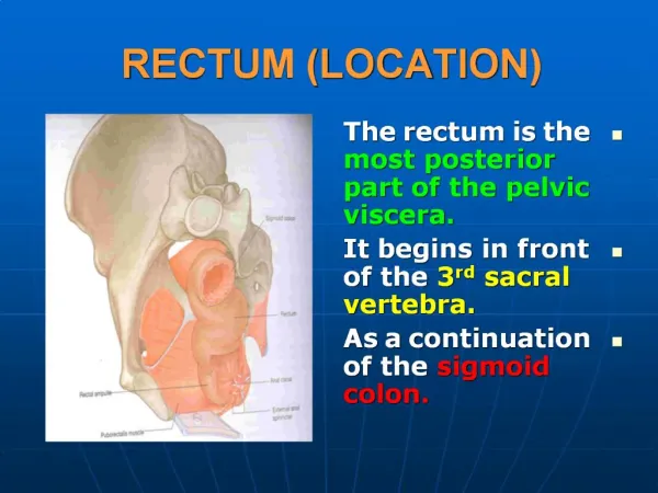
Colon and Rectum. Anatomic Considerations and Patterns of Spread. Rectum . 12 to 15 cm in length from the rectosigmoid junction to the puborectalis ring upper third middle third (posterior border of the rectouterine pouch or rectovesical space) lowest third no serosal barrier.

Colon and Rectum
E N D
Presentation Transcript
Rectum • 12 to 15 cm in length • from the rectosigmoid junction to the puborectalis ring • upper third • middle third (posterior border of the rectouterine pouch or rectovesical space) • lowest third no serosal barrier
Colonic nodal drainage • consists of pericolic nodes and nodes in association with the vascular supply to the colon (i.e., mesenteric nodes • rectal nodal drainage include the perirectal, presacral, and internal iliac nodes .
Epidemiology, Molecular Cascade, Risk Factors, Hereditary Disease, and Clinical Presentation • The median age is in the seventh decade • chemical carcinogens • Environmental and dietary factors
Factors shown to increase the risk • include increasing age, • male sex, rectal cancer • excessive alcohol use • family history of colorectal cancer, • increasing height, • increasing body mass index, • processed meat intake • low folate consumption. • consumption of fruits and vegetables ? • The role of chemopreventive agents (carotenoids, aspirin, and other nonsteroidal anti-inflammatory drugs) ?
Biologic and genetic pathways of development of colorectal cancer • proto-oncogenes (mutations in the ras proto-oncogene.) • tumor suppressor genes (inactivation adenomatous polyposis coli (APC) and P53)
:microsatellite “instability”Biology • The majority of HNPCCas well as a minority of sporadic colorectal cancers harbor microsatellite “instability.” • mutations in genes encoding enzymes that repair DNA replication errors • Studies have suggested that patients with tumors possessing a high frequency of microsatellite instability have a more favorable outcome and develop fewer metastases
Colorectal cancer • minimal or no symptoms • Nonspecific bowel habits, weakness, intermittent abdominal pain, nausea, and vomiting. The persistence of such symptoms as well as any evidence of iron deficiency anemia should be investigated
Clinical Presentation • right colon exophytic ,iron deficiency anemia • left colon and sigmoid colon deeply invasive, annular, and accompanied by obstruction and rectal bleeding • Rectal cancer frequently results in bleeding and alterations in bowel habits.
FIGURE 58.1. Idealized depiction of peritoneal relationships in the colon and rectum. The transverse and sigmoid colon are intraperitoneal, with a complete peritoneal covering (serosa) and mesentery. The ascending and descending colon are retroperitoneal, lack a true mesentery, and usually do not have a peritoneal covering posteriorly or laterally. The upper rectum begins above the peritoneal reflection and has peritoneum anteriorly and laterally. The lower half to two thirds of the rectum is below the peritoneal reflection (infraperitoneal).
Prevention and Early Detection • Neoplastic polyps are precursors of colon cancers • Most colorectal cancers arise from pre-existing polyps • Patients with neoplastic polyps should be considered at high risk for large bowel cancer, and polypectomy may reduce this risk. With the availability of the flexible colonoscope and endoscopic polypectomy, polyps can be removed at a premalignant stage and patients followed closely.
The goal of screening is to detect preinvasive polyps or early invasive cancer. The presence of polyps increases the risk for cancer to approximately 15%. Data from programs in which proctoscopy is performed annually suggest that routinely scheduled polypectomy reduces the development of subsequent bowel cancer by 80% or more .
The American Cancer Society has recommended screening should begin at age 50 in the average risk patient by either: • Annual fecal occult blood test and/or flexible sigmoidoscopy every 5 years, • Double contrast barium enema every 5 years • Colonoscopy every 10 years
patients at high risk • adenomatous polyps • history of colorectal cancer • first-degree relative with colorectal cancer or adenomas • inflammatory bowel disease • family history or genetic testing
Intensive surveillance is recommended for patients at high risk • Computed tomography (CT) colonography • genetic fecal testing are being studied Although screening methods can detect colorectal cancer at an early stage, <40% of patients are diagnosed with early disease, likely reflecting low rates of disease awareness as well as the infrequency of screening in eligible candidates
Pathology • (>90%) adenocarcinomas • Scc, carcinoid, leiomyosarcoma, and lymphoma. • Most grading systems classify adenocarcinoma as well, either moderately or poorly differentiated.
Pathways of Spread • invade from mucosa through the bowel wall and beyond, with involvement of lymphatic channels and lymph nodes. • Hematogenous spread can occur, primarily to the lung and liver. • There is little propensity for colon cancer to spread longitudinally within the bowel wall, in contrast to esophageal or gastric cancers
Patient Evaluation/Staging • Pretreatment evaluation • pathological confirmation • colonoscopy ( synchronous primaries occurring in 3% to 5%) • CBC with LFT and CEA • abdominal and pelvic CT scan • CXR • (PET) scan • (MRI), • Ultrasound
(PET) scan • در مرحله بندی قبل عمل ارزشمند نیست اما در مرحله بندی بعد درمان ،عود یا شک به عود مفید است دویتا • در موارد الیگومتاستاز کاندید درمان شفاء دهند ه مفید است.پرز
Prognostic factors • depth of tumor invasion into and beyond the bowel wall • the number of involved regional lymph nodes • presence or absence of distant metastases • The tumor, node, metastasis (TNM) system of the American Joint Committee on Cancer can be used as a clinical (preoperative) or postoperative staging system
Colorectal Tumor, Node, Metastasis Staging, 2002 • Tis Carcinoma in situ: Intraepithelial or invasion of lamina propria • T1 Tumor invades submucosa • T2 Tumor invades muscularis propria • T3 Tumor invades through the muscularis propria into the subserosa, or into non-peritonealized pericolic or perirectal tissues • T4 Tumor directly invades other organs or structuresa and/or perforates visceral peritoneum (includes invasion of other segments of colon)
Direct invasion in T4 includes invasion of other segments of the colorectum by way of the serosa; for example, invasion of the sigmoid by a carcinoma of the cecum. Tumor that is adherent to other organs or structures, macroscopically, is classified as T4. However, if no tumor is present in the adhesion, microscopically, the classification should be pT3
N1 Metastasis in one to three regional lymph nodes • N2 Metastasis in four or more regional lymph nodes (Tumor nodules in the pericolonic adipose tissue without evidence of residual lymph node are classified as a regional lymph node metastases)
Therapy of Colon CancerSurgery • based on the anatomy and mechanisms by which this disease spreads. • avoid cutting across tumor in intramural lymphatics, • sufficient lengths of bowel must be resected
Resection results in excellent cure rates average 5-year survival • 97% for T1N0 • 85% to 90% for T2N0 • 65% to 75% for T3–T4N0 • 50% (T3N+) and 35% (T4N+)
Adjuvant Chemotherapy • addition of 5FU (5-fluorouacil) and leucovorin improves survival for resected stage III patients • Capecitabine, an oral 5-FU prodrug, has demonstrated similar overall and disease free survival rates to 5-FU/leucovorin in patients with resected stage III colon cancer • 5-FU/leucororin/oxaliplatin in resected stage II or III colon cancer patients showed improved disease-free • FU/LV/oxaliplatin as the new standard chemotherapeutic regimen in the adjuvant treatment of completely resected, high-risk colon cancer • The efficacy of agents such as bevacizumab and cetuximab as adjuvant therapy is being investigated
Adjuvant Irradiation with or without Concurrent Chemotherapy • The potential indications for adjuvant radiation therapy in colon cancer are based on analyses of patterns of failure following resection • local failure in colon cancer also depends on anatomic origin • ascending and descending colon • mid-sigmoid and mid-transverse colon • cecal, hepatic/splenic flexure, and proximal/distal sigmoid tumors are variable depending on the amount of mesentery present, tumor extension, and the adequacy of radial margins. • When colon cancers adhere to or invade adjacent structures, local failure rates exceed 30% following surgery alone.
In summary, local failure occurs in patients with colonic tumors where there are anatomic constraints on radial resection margins, including tumors adherent to or invading adjacent structures.
To summarize, these studies have suggested that operative bed failures in high-risk patients undergoing resection alone are at least 30%, and that the risk of local failure is reduced by the administration of adjuvant radiation therapy. • Irradiated patients included those with T4N0/N+, T3N+ disease (excluding mid-sigmoid and mid-transverse colon) and T3N0 patients with margins of <1 cm.
((T3–4N0/N+ tumorscompared with CHRT surgery alone • radiation, with concurrent chemotherapy, usually with bolus 5-FU (500 mg/m2/d) for 3 consecutive days during the first and last weeks of radiation therapy. Radiation treatment to treat the tumor bed with an approximate 3- to 5-cm margin to a total dose of 45 Gy, • Draining nodes were included if thought to be at high risk for involvement. Local control rates in T4N0 and T4N+ patients treated with radiation therapy were 93% and 72%, respectively versus 69% and 47%, respectively in patients undergoing surgery alone. Similarly, relapse-free survivals were 79% and 53%, respectively in T4N0/T4N+ patients undergoing adjuvant radiation, versus 63% and 38%, respectively undergoing surgery alone. • No significant outcome differences were observed in patients with T3N0 and T3N+ lesions; however, there may be an element of selection bias in the radiation group given most patients were referred because of concerns of adequacy of local control following surgery alone.
CHRT در کنسر کولون • The rate of acute enteritis in patients receiving irradiation and 5-FU was 16% versus 4% in patients undergoing irradiation only. • Late bowel complication rates were not increased by concomitant 5-FU administration. • with increasing numbers of nodes involved, survival steadily decreased • There was a trend toward improved local control in patientsreceiving 5-FU
اندیکاسیون CHRT در کنسر کولون • T4 tumors • margin positive resection • abscess/fistula formation
FIGURE 58.2. Idealized postoperative anteroposterior-posteroanterior irradiation fields of extrapelvic colon cancer (tumor bed and nodal regions). If treated preoperatively, lateral fields could be added based on imaging with computed tomography of the abdomen and colon radiograph. A: Para-aortic nodes are at risk, in addition to tumor bed, due to tumor adherence to posterior abdominal wall with descending colon cancer. B: External and common iliac nodes are at risk, in addition to tumor bed, from proximal ascending colon cancer.
local control rates in patients undergoing intraoperative boost were 89% compared to 18% undergoing external irradiation alone • 5-year survival margin negative resection (66%) microscopic residual (47%) gross residual (23%). • patients undergoing intraoperative boost demonstrated improved survival (76% vs. 26%).
A study from the University of Florida of patients with locally advanced but completely resected colon cancers receiving adjuvant radiation reported a local control rate of 88%, similar to the 90% reported at the Mayo Clinic in patients who had completely resected tumors. • there appeared to be a dose-response relationship to local control. The 5-year rate of local control was 96% for patients receiving 50 to 55 Gy versus 76% for patients receiving <50 Gy
To assess whether the addition of radiation therapy to adjuvant chemotherapy would result in superior survival and local regional failure rates in resected, high-risk colon cancer patients, the U.S. Intergroup initiated a randomized prospective trial in 1992 (103). In this trial, patients with resected colon cancer were randomized to postoperative irradiation with 5-FU and levamisole or 5-FU and levamisole alone. Eligibility criteria included margin negative tumors with adherence to or invasion of surrounding structures (i.e., T4N0 or N+ disease, excluding peritoneal invasion) or tumors arising in the ascending or descending colon with metastatic regional nodes (T3N+). Patients were randomized to receive (a) weekly 5-FU combined with levamisole for 12 months' duration or (b) 5-FU and levamisole for 12 months with combined radiation therapy and chemotherapy beginning 1 month after the first 5-FU administration. The recommended total radiation dose was 45 Gy in 25 fractions over 5 weeks with an optional 5.4 Gy boost. • No definitive conclusions can be made regarding the efficacy of postoperative irradiation with 5-FU and levamisole based on this underpowered study with many flaws; however, this study provides no data supporting its routine use.
IOERT • local failure 11% receiving IOERT plus EBRT versus 82% EBRT only • 5-year survival 76% for patients receiving IOERT 26% for patients receiving EBRT alone]
metastatic disease • 5-FU–based chemo-therapy is usually administered. • Recent prospective randomized trials have shown that multiple agents improve survival in patients with metastatic colorectal cancer.
irinotecan, 5-FU, leucovorin, oxaliplatin • Varying combinations of these drugs as well as other novel agents remain the focus of ongoing investigation in both the metastatic and nonmetastatic setting.
Palliative CHRT • Palliative irradiation, sometimes in combination with 5-FU–based chemotherapy, is considered for patients with specific symptoms referable to metastatic disease—brain, bone, and other sites. • The combination of radiation therapy and newer agents (capecitabine, irinotecan, oxaliplatin, bevacizumab, cetuximab) remains investigational
Techniques of Irradiation • Field arrangement will vary depending on the site of the primary disease as well as areas judged to be at high risk for local recurrence • Patient positioning (supine, prone, decubitus) should be considered in planning. • the right or left decubitus position for at least a portion of their treatment, allowing displacement of the small bowel away from the treatment field. • Immobilization devices may improve reproducibility. • It may be useful to compare films in both the decubitus and supine positions to determine the actual amount of small bowel displacement. • CT-based planning may facilitate defining the tumor bed, determining beam orientation, as well as estimating the volume of small bowel included within the treatment fields.
Techniques of Irradiation • total radiation dose 45 Gy in 25 fractions of 1.8 Gy per fraction (primary tumor and at-risk tissues) Reduced fields to 50 Gy T4 tumors( a total dose of 54 to 60 Gy)