Wound care
Wound care. Professor Magdy Amin RIAD Professor of Otolaryngology. Ain shames University Senior Lecturer in Otolaryngology University of Dundee. There are four basic tissues in the body:. 1) epithelium; 2) Connective tissues, including blood, bone and cartilage; 3) muscle tissue;

Wound care
E N D
Presentation Transcript
Wound care Professor Magdy Amin RIAD Professor of Otolaryngology. Ain shames University Senior Lecturer in Otolaryngology University of Dundee
There are four basic tissues in thebody: 1) epithelium; 2) Connective tissues, including blood, bone and cartilage; 3) muscle tissue; 4) nerve tissue.
The parameters for measuring thestrength of normal body tissue are: • Tensile Strength • Breaking Strength • Burst Strength
Tensile Strength The load per cross-sectional area unit at the point of rupture, relating to the nature of the material rather than its thickness.
Breaking Strength • The load required to break a wound regardless of its dimension, the more clinically significant measurement.
Burst Strength • The amount of pressure needed to rupture a viscus, or large interior organ.
RECOVERY OFTENSILE STRENGTH • As collagen accumulates during the reparative phase, strength increases rapidly but it is many months before a plateau is reached. • Until this time, the wound requires extrinsic support from the method used to bring it together – usually sutures.
RECOVERY OFTENSILE STRENGTH • While skin and fascia are the strongest tissues in the body, they regain tensile strength slowly during the healing process. • The stomach and small intestine, on the other hand, are composed of much weaker tissue but heal rapidly.
PATIENT FACTORS THATAFFECT WOUND HEALING • Age • Weight • Nutritional status • Dehydration • Inadequate blood supply to the wound • Chronic disease • Radiation therapy
Surgical Factors that affect wound healing • The length and direction of the incision • Dissection technique • Tissue handling • Hemostasis • Maintaining moisture in tissues • Removal of necrotic tissue and foreign materials
THE LENGTH ANDDIRECTION OF THEINCISION • The direction in which wounds naturally heal is from side-to- side, not end-to-end. • The arrangement of tissue fibers in the area to be dissected will vary with tissue type. • The best cosmetic results may be achieved when incisions are made parallel to the direction of the tissue fibers.
DISSECTION TECHNIQUE When incising tissue, a clean incision should be made through the skin with one stroke of evenly applied pressure on the scalpel. Sharp dissection should be used to cut through remaining tissues. The surgeon must preserve the integrity of as many of the underlying nerves, blood vessels, and muscles as possible.
TISSUE HANDLING • Keeping tissue trauma to a minimum promotes faster healing. • Throughout the operative procedure, the surgeon must handle all tissues very gently and as little as possible. • Retractors should be placed with care to avoid excessive pressure, • since tension can cause serious complications: impaired blood and lymph flow, • altering of the local physiological state of the wound, and predisposition to microbial colonization.
HEMOSTASIS • Achieving complete hemostasis before wound closure will • prevent formation of postoperative hematomas. • Collections of blood (hematomas) or fluid (seromas) in the incision can prevent the direct apposition of tissue. • these collections provide an ideal culture medium for microbial • growth and can lead to serious infection. • When clamping or ligating avoid excessive tissue damage. • Mass ligation that involves large areas of tissue may • produce necrosis.
MAINTAINING MOISTUREIN TISSUES • During long procedures, periodically irrigate the wound with warm saline solution, • or cover exposed surfaces with saline-moistened sponges to prevent tissues from drying out.
REMOVAL OF NECROTIC TISSUE AND FOREIGN MATERIALS • Adequate debridement of all devitalized tissue and removal of inflicted foreign materials are essential to healing
CHOICE OF CLOSURE MATERIALS • Cellular response to closure materials • Closing tension • Postoperative distraction forces • Immobilization • Elimination of dead space in the wound
CLASSIFICATION OF OPERATIONClass Definition • Clean Operations in which no inflammation is encountered . The respiratory, alimentary or genitourinary tracts are not entered. There is no break in aseptic operating theatre technique.
CLASSIFICATION OF OPERATIONClass Definition Clean-contaminated Operations in which the respiratory, alimentary or genitourinary tracts are entered but without significant spillage.
CLASSIFICATION OF OPERATIONClass Definition Contaminated Operations where acute inflammation (without pus) is encountered. or where there is visible contamination of the wound. Examples include gross spillage from a hollow viscus during the operation or compound/open injuries operated on within four hours.
CLASSIFICATION OF OPERATIONClass Definition Dirty Operations: In the presence of pus. where there is a previously perforated hollow viscus, or compound/open injuries more than four hours old.
HEALING BYPRIMARY INTENTION • Inflammatory (preparative) • Proliferative • Remodelling • Overlapping phases.
The inflammatoryphase • lasts for three to five days. • Characterized by edema, erythema, heat and pain. • Immediately following the injury hemostasis occurs. • Various cells such as leukocytes and macrophages migrate to the injury to stimulate coagulation, vasodilation and increased capillary permeability.
The inflammatoryphase • Neutrophils migrate to the injury to control bacteria. • Platelets release cytokines (proteins such as growth factors) that act as cellular mediators (or messengers). • These substances stimulate and contro linflammation and the formation and growth of new epidermis, tissue granulation, blood vessels and scar tissue.
GROWTH FACTORS FOUNDIN WOUND FLUID • Platelet-derived growth factor (PDGF) • Transforming growth factor alpha and beta (TGFa and TGFb) • Basic fibroblast growth factor (bFGF) • Epidermal growth factor (EGF) • Insulin growth factor 1 (IGF-1)
The proliferative (granulation)phase • lasts several weeks and is likewise controlled by growth factors. • Granulation tissue formed and collagen, which eventually gives tissue tensile strength and structure, is produced.
The proliferative (granulation)phase Epithelialization • Epithelialization phase, epidermal cells close the wound and form a complete scar. • Regulated amounts of matrix metalloproteinases (MMPs) mediate many of the cellular processes that occur during epithelialization
The final remodeling (maturation) phase • The scar tissue matures and becomes stronger through matrix degradation and formation. • This phase, which can last for six months to several years, is also controlled by growth factors and results in scar tissue that has about 80% of the tensile strength of uninjured skin.
How chronic woundsdiffer from acute wounds • In acute wounds, the body advances through these phases in an orderly and timely manner. • When the body does not progress accordingly the injury is known as a “chronic wound.”
How chronic woundsdiffer from acute wounds • Pressure, trauma,edema, abnormal or excessive bacterial presence and decreased oxygen are local factors that contribute to delayed wound healing. • Age, chronic disease and nutritional status are examples of systemic factors that may impede wound healing.
How chronic woundsdiffer from acute wounds • These factors may affect the quantity, timing and activity of the cytokines, growth factors and MMPs. • Certain growth factors, including PDGF, EGF and TGFb, are found in decreased levels in chronic wounds compared to acute wounds. • In some cases, these substances may be present, but not available to the wound for healing. • In addition, bacteria found in chronic wounds produce high levels of enzymes and MMPs. • These substances degrade the cytokines, eliminating the mediators of cellular processes that are critical to wound healing.
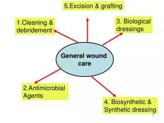
Three traditional cornerstonesconcepts of wound management • Moist wound healing. • Debridement. • Management of the wound fluid (exudate)
Moist woundhealing • occlusive, semiocclusive and hydrocolloid dressings introduced to maintain a moist environment in wounds. • increase epithelialization, promote dermal matrix synthesis and improve the patient’s comfort.
The Exudate • The exudate of chronic wounds has been found to inhibit wound healing. • Studies have demonstrated that chronic wound fluid can inhibit the production of key woundhealing cells (e.g., keratinocytes, fibroblasts and vascular endothelial cells). • Bacterial exudate also contains increased levels of MMPs, which can inhibit wound healing by degrading growth factors or reducing the number of active growth factor receptor sites on the cells. • Removing or controlling exudate can lead to improved wound healing.
The Exudate • Compression continues to be one of the more common methods of exudate management. • Negative pressure devices and vacuum-assisted devices, are now being used to improve wound healing. • Drains are important in preventing exudate from accumulating in wound site.
HEALING BYSECOND INTENTION • Healing by second intention is caused by infection, excessive trauma, tissue loss, or imprecise approximation of tissue. • Granulation tissue forms and contains myofibroblasts. These help to close the wound by contraction. • This process is much slower than primary intention healing. • Excessive granulation tissue may build up and require treatment if it protrudes above the surface of the wound, preventing epithelialization.
DELAYED PRIMARYCLOSURE • method of management of contaminated, as well as dirty and infected traumatic wounds with extensive tissue loss and a high risk of infection. • The surgeon usually treats these injuries by debridement of nonviable tissues and leaves the wound open, inserting gauze packing which is changed twice a day.
DELAYED PRIMARYCLOSURE • Patients sedation or a return to the operating room with general anesthesia generally is only required in the case of large, complex wounds. • Wound approximation using adhesive strips, previously placed but untied sutures, staples after achieving local anesthesia can occur within 3-5 days if the wound demonstrates no evidence of infection and the appearance of red granulation tissue.
DELAYED PRIMARYCLOSURE • Should this not occur, the wound is allowed to heal by secondary intention.
THE WOUND • Injury to any of the tissues of the body, especially that caused by physical means and with interruption of continuity is defined as a wound.
Wound healing is a natural and spontaneous phenomenon. • When tissue has been disrupted so severely that it cannot heal naturally : • dead tissue and foreign bodies must be removed, • infection treated, • and the tissue must be held in apposition
until the healing process provides the wound with sufficient strength to withstand stress without mechanical support. • A wound may be approximated with sutures, staples, clips, skin closure strips, or topical adhesives.
There are four basic tissues in thebody: • epithelium; • connective tissues, including blood, bone and Cartilage 3) muscle tissue 4) nerve tissue. The choice of wound closure materials and the techniques of using the are prime factors in the restoration of continuity and tensile strength to the injured tissues during the healing process.