Download
1 / 31
680 likes | 2.43k Views
Palliative Wound Care. PCQN May 15th Susan Barbour RN MS FNP CWON ACHPN. Objectives. Palliative wound care Pressure Ulcers Skin Failure Fungating wound management Complex abdominal wounds. Ostomy Care. Wounds & EOL. Types of wounds at EOL Pressure ulcers – most common Skin Failure

E N D
Palliative Wound Care PCQN May 15th Susan Barbour RN MS FNP CWON ACHPN
Objectives • Palliative wound care • Pressure Ulcers • Skin Failure • Fungatingwound management • Complex abdominal wounds
Wounds & EOL • Types of wounds at EOL • Pressure ulcers – most common • Skin Failure • Malignant fungating wounds in advanced cancer • Complex Surgical wounds • Any other chronic wound – venous, arterial, DM… • Symptom management in wounds at EOL (pain, exudate, odor, bleeding, infection) • Wound Treatment • Principles • Dressings
Wound Assessment • Approach patient with sensitivity as often self conscious, embarrassed, upsetting • Get detailed history of wound • Other providers involved? Home Health Nursing • History of impact on patient and family • What is the most bothersome about the wound? Can use ‘bother scale’ 0-10 • Caregiver burden? • Difficulties with providing care (location, cost) • Determine etiology. May be multifactorial • Need for specialized supplies (air mattress) • Exam (pre-medicate if necessary) : Location, size, exudate, surrounding skin, odor, pain, amount of devitalized tissue, bleeding
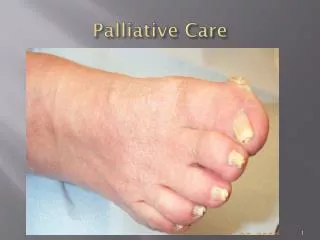
Pressure Ulcers • 6 stages • Only stage pressure ulcers • Common at EOL • GOC determine treatment • If present on admission, address possible family guilt • State reportable if full thickness ulcer acquired during hospitalization • Evolve predictably but often delay in appearance on skin • Not all pressure ulcers are avoidable • Skin Failure/Kennedy Ulcer
How long does it take to get a pressure ulcer? Paraplegic patient who spent several hours on a gurney before operation. Evolved to Stage IV ulcer.
Full thickness OR acquired pressure ulcer Post-operative day one after valve surgery. SuspectedDeep tissue injury. Now unstageable 30 days later. Stage IV pressure ulcer after debridement
Stage I Pressure Ulcer • Intact skin with non-blanchable redness of a localized area usually over a bony prominence. Darkly pigmented skin may not have visible blanching; its color may differ from the surrounding area. • The area may be painful, firm, soft, warmer or cooler as compared to adjacent tissue. Stage I may be difficult to detect in individuals with dark skin tones.
Assessing for early pressure ulcers in dark skinned individuals • Cannot rely on changes in skin color. • Must be extra vigilant about assessing at-risk individuals • Patients may complain of pain or itching in the area where a pressure ulcer is evolving • Feel the skin for changes in temperature and consistency (note if firm, mushy, boggy, warmer or cooler) as compared to adjacent tissue.
Stage II Pressure Ulcer • Partial thickness loss of dermis presenting as a shallow open ulcer with a red pink wound bed, without slough. May also present as an intact or open/ruptured serum-filled blister. • Presents as a shiny or dry shallow ulcer without slough or bruising.* This stage should not be used to describe skin tears, tape burns, perineal dermatitis, maceration, or excoriation. * Bruising indicates suspected deep tissue injury.
Stage III Pressure Ulcer • Full thickness tissue loss. Subcutaneous fat may be visible but bone, tendon or muscle are NOT exposed. Slough may be present but does not obscure the depth of tissue loss. May include undermining and tunneling. • The depth of a stage III pressure ulcer varies by anatomical location. The bridge of the nose, ear, occiput, and malleolus do not have subcutaneous tissue and stage III ulcers can be shallow. In contrast, areas of significant adiposity can develop extremely deep stage III ulcers. Bone/tendon is not visible or directly palpable.
Stage IV Pressure Ulcer • Full thickness tissue loss with exposed bone, tendon or muscle. Slough or eschar may be present on some parts of the wound bed. Often includes undermining and tunneling. • The depth of a stage IV ulcer varies by anatomical location. Stage IV ulcers can extend into muscle and/or supporting structures (fascia, tendon or joint capsule) making osteomyelitis possible. Exposed bone/tendon is visible or directly palpable.
Unstageable • Full thickness tissue loss in which the base of the ulcer is covered by slough (yellow, tan, gray, green or brown) and/or eschar (tan, brown or black) in the wound bed. • Until enough slough and/or eschar is removed to expose the base of the wound, the true depth, and therefore stage, cannot be determined. Stable (dry, adherent, intact without erythema or fluctuance) eschar on the heels serves as “the body’s natural (biological) cover” and should not be removed.
Suspected Deep Tissue Injury • Purple or maroon localized area of discolored intact skin or blood-filled blister due to damage of underlying soft tissue from pressure and/or shear. The area may be preceded by tissue that is painful, firm, mushy, boggy, warmer or cooler as compared to adjacent tissue. • Deep tissue injury may be difficult to detect in individuals with dark skin tones. Evolution may include a thin blister over a dark wound bed. The wound may further evolve and become covered by thin eschar. Evolution may be rapid exposing additional layers of tissue even with optimal treatment.
Skin Failure at end of Life “an event in which the skin and underlying tissue die due to hypoperfusion that occurs concurrent with severe dysfunction or failure of other organ systems. Skin failure can be categorized as acute, chronic, or end stage. Pressure ulcers, a type of skin death, frequently occur in persons with a heavy disease burden, especially those at or near the end of life, despite good care” Diane Langemo RN PhD
Pressure Ulcer prevention • Repositioning – in EOL patients this should be discussed with the family • Pressure redistribution surface – Medicare guidelines • Risk assessment scales – Braden scale • Risk factors – early identification and intervention • ED early assessment & intervention • Education to patient and family
Malignant Fungating Wounds • May occur in up to 5% in pts with cancer • 10% of patients with metastatic disease • Locally advanced, metastatic, recurrent • For women • 70.7% breast carcinoma • 12% melanoma • For men • 32.3% melanoma • 11.8% lung cancer • 11% colorectal cancer • Other locations • Head and neck, ovarian, GU
Symptom Management: Wound Pain • Current analgesia and effect • Determine baseline usual pain and what causes increased pain • Emotional pain (anxiety, anticipatory pain) • Determine strategies that minimize pain • Often due to dressings that ‘stick’ to the tissue – try Vaseline gauze or similar • More frequent dressing changes avoid sticking • Warm versus cold cleansing solutions • Moist wound healing • Leaving tissue open to air usually more painful/letting tissue ‘dry out’ • Pre-medicate before dressing changes • Tell pt to STOP (time out) dressing change procedure as needed • Have them remove dressings • Topical opiods added to hydrogel • Palliative amputation
Odor Assessment Necrotic tissue, infection, bacterial load • Assessment: • “Is there any odor to the wound or the drainage?” • “Is the odor a problem?” • “What have you already tried?” “Did it work?” • “Does the odor keep you from doing things you enjoy doing?” • When is the odor problematic? • Delayed dressing change • Recent dressing change
Malignant Wounds OdorTreatment • If possible, remove loose necrotic debris & tissue • Wound cleansing – incontinence cleansers, showers or irrigation • Apply topical metronidazole (Metrogel or solution made by crushing metronidazole tablets in sterile water) • Dilute Dakin’s solution (0.05%) • Silvadene • Charcoal dressings, topical yogurt, • Occlusive dressing to trap odors • Diapers, panty liners • Kitty litter or charcoal briquettes under the bed • Odor eliminating sprays, aromatherapy
Exudate management • Amount of drainage? • What has worked to catch it? • Is it leaking onto their clothing? • Drainage management • Diapers, pads, alginates, foam • Avoid tape (often previously radiated tissue) and use alternative securing system (undershirt, briefs, ace wrap, flexinet)
Bleeding • When does the wound bleed? • How much and is it difficult to stop the bleeding? Transfusions? • Are they curtailing their activities because of the bleeding? • Often dressing related • Care on removing dressings. Soak if necessary. • Maintain moist wound environment • Gentle cleansing • Use of non fibrous dressing materials • More frequent dressing changes
Malignant WoundsPsychological burden and QOL • Determine impact on patient and family • Grief, anxiety, embarrassment, stigma, withdrawal • Anal/rectal tumors makes difficult to sit • Extremity swelling • Identify strategies to minimize impact • Techniques to cover tumors • Openly discuss issues
Types of Dressings • The type of dressing changes as the tumor grows and conditions change • Amount of exudate • Size of wound • Condition of tissue • Location of patient • Ensure dressing is a good match for the patient and their circumstances • Insurance coverage • Ease of use and understanding of application • Pain of dressing change • Level of “skilled need”
Hydrogels • Amorphous gels, sheets and impregnated gauze • Water or glycerin based • Minimal absorbency • Minimal pain • Most case studies on using topical morphine used Intra-Site Gel as base for morphine
Foams • Polyurethane foam • Variable thickness • Non adhesive wound contact surface • May have surrounding adhesive to secure • High absorbency of exudate
Alginate dressings • Calcium alginates composed of non-woven fibers of a cellulose-like polysaccharide derived from the calcium salt of alginic acid (seaweed) • Available in varying forms, ropes for packing cavities, ribbons for narrow wounds or sinuses, or flat pads. • Absorbent dressings that minimize pain and can allow for decreased frequency of dressing. • Most require secondary dressing • Common examples: Kaltostat, Curasorb, Restore, Sorbsan • WoundSource 2009