Download
1 / 9
310 likes | 5.47k Views
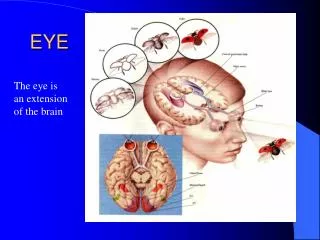
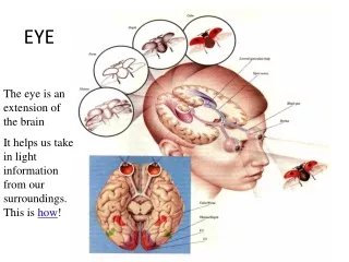
Eye Examination. Heather Nelson, RN. Inspection of the Eye. eyebrows---size, extension, and texture of hair eyelids---color, edema, lesions, adequate muscle control, eyelashes, ability to close completely and open widely sclera---color, vascular pattern, should be white.

E N D
Eye Examination Heather Nelson, RN
Inspection of the Eye • eyebrows---size, extension, and texture of hair • eyelids---color, edema, lesions, adequate muscle control, eyelashes, ability to close completely and open widely • sclera---color, vascular pattern, should be white
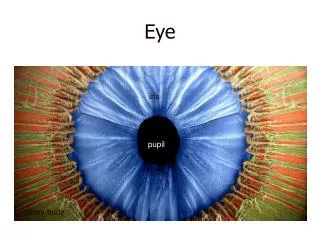
Inspection of the Eye • Cornea---examine for clarity by shining a light on it. Blood vessels should not be present. • Iris---Should be round, regular, and equal in size. Note color. • Conjunctiva---usually free of erythema and exudate. Only inspect upper when foreign object may be present.
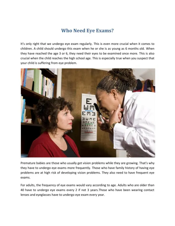
Eyes • need Snellen eye chart or Rosenbaum pocket vision card • need opthalmoscope • check for color blindness • check visual acquity---far at 20 feet, near at 14 inches
Pupils • size, shape, symmetry • reaction to light • accommodation
Visual Fields by Confrontation • do both eyes together • wiggle fingers at side of patient’s head • repeat 2-3 times to test both temporal fields • test eyes separately if abnormality is suspected
Extraocular Muscles • Have patient watch your finger as it moves through the six cardinal fields of gaze. • Test for convergence. • Observe for sustained nystagmus.
Opthalmoscopic Exam • Darken room. • Start at 0 diopters. • Use plain white circle of light. • Ask pt to focus on spot on wall. • Find red reflex from 2 feet away. Caused by light illuminating the retina. • Follow in on 15 degrees. • Adjust diopters to focus. • Look at vascular supply of the retina. • Follow blood vessels to optic disc. Disc margin should be sharp and well-defined.
What does 20/20 mean? • Visual acuity is recorded as a fraction in which the numerator indicates the distance of the patient from the chart and the denominator indicates the distance at which the average eye can read the line. • Thus 20/20 means that the patient can read at 20 feet what the average person can read at 20 feet. • Vision not correctable to better than 20/200 is considered legal blindness.