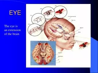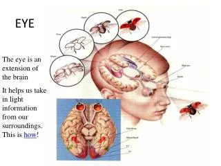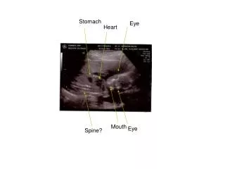
Eye
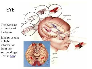
Eye. iris. pupil. ciliary body. Eyeball. FIBROUS. VASCULAR. NEURAL. Eyeball. Eyeball in LM. posterior segment. anterior segment. Eyeball. Three layers of the eye Multilayer inner retina Middle choroid – pigmented, vascular Outer sclera – dense fibrous c . t.

Eye
E N D
Presentation Transcript
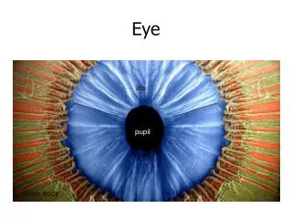
Eye iris pupil ciliary body
Eyeball FIBROUS VASCULAR NEURAL
Eyeball in LM posterior segment anterior segment
Eyeball • Three layers of the eye • Multilayer inner retina • Middle choroid – pigmented, vascular • Outer sclera – dense fibrous c . t.
Fibrous tunic - tunica externa oculi Cornea Sclera
Cornea • Stratified squamous epithelium • Bowman´s membrane-anterior limiting lamina • Substantia propria cornae • 200 - 250 layers of regularly organized collagen fibrils • fibrocytes /keratocytes/ • Descemet´s membrane-posterior limiting lamina • the basement membrane of the posterior endothelium • Posterior endothelium • simple squamous epithelium
Vascular tunic - tunica media oculi • Choroid - loose c.t. with network of blood vessels, numerous pigment cells • Ciliary body - loose c.t. with smooth muscle cells – musculus ciliaris /accomodation/ - ciliary processes – generate aqueous humor • Iris - central opening of the iris - the pupil ch c i
Choroid • Lamina suprachoroidea /lamina fusca sclerae/ • Lamina vasculosa • Lamina chorocapillaris • Lamina vitrea /Bruch´s membrane/ sclera choroid retina
Ciliary body - structure ciliary processes m. ciliaris ciliary epithelium - outer cell layer is pigmented, whereas the inner cell layer is nonpigmented /pars caeca retinae/
Ciliary body Two functions • - accomodation • - production of the aqueous humor ACCOMODATION • from the ciliary processes extend fibers towards the lens - fibres are called zonular fibres • contraction of the m. ciliaris reduces the tension of the zonule fibres and result in a thickening of the lens which focusses on close objects - accomodation
Iris • Anterior epithelium • discontinued layer • Anterior border layer • pigment cells • Stroma iridis • gelatinous c.t., numerous pigment cells • surrounds the pupil are smooth muscle cells which form the annular sphincter pupillae muscle • Posterior border layer • m. dilator pupillae /myoepithelial cells/ • Posterior epithelium • one layer pigmented cells • pars iridica retinae colour of the eyes
Retina ora serrata
Retina (neural tunic) – cell types • Pigment cells • Neurons • 1st neuron = rod cells and cone cells • 2nd neuron = bipolar cells • 3rd neuron = ganglion cells (multipolar) • interneurons • horizontal cells • amacrine cells • Neuroglia • Müller cells – occupy practicly the entire retina, part between outer and inner limiting membrane (which are formed by their cell bases)
Retina inner limiting membrane 10 layers
Retina light
Retina - fovea centralis maximal visual acuity only cone cells
Retina - discus nervi optici optic disc - blind spot sclera
Refractive media • Cornea • Aqueous humor • Lens • Vitreous body Refractive media are characterized by high transparency and refractivity.
Lens biconvex body The lens fibers are highly specialized cells that differentiate from lens epithelium cells.
Lens • lens capsule • is generated by the cells of the subcapsular epithelium • subcapsular epithelium • anterior and lateral parts→stayes • posterior part→lens fibers • lens fibres • very long (up to 12 mm), hexagonal cells, form the body of the lens • lens fibres are nucleated in the soft, outer cortex of the lens • lens fibres located deeper loose their nuclei and become part of the harder nucleus of the lens
Fasciculus opticus – optic nerve • is surrounded by the three meninges • c.t. septa, which arise from the pia mater, separate the fibre bundles in the optic nerve • the axons in the optic nerve are supported by astrocytes and oligodendrocytes, microglia is also present
Eyelid cutaneous part tarsal plate conjunctival part Meibomian glands – tarsal /sebaceae/ Zeiss glands /gl. sebaceae ciliares/ Moll glands /gl. sudoriferae ciliares – apocrine/
Eyelid /palpebra oculi/ tarsal plate and tarsal Meibomian glands /sebaceae/
Conjunctiva stratified columnar epithelium with goblet cells lamina propria mucosae
Lacrimal apparatus • lacrimal gland • compound tubuloalveolar gland producing a lysosyme-rich serous fluid • lacrimal canaliculi • superior • inferior • lenghth: 8 mm, lined with s.s.epi • lacrimal sac • nasolacrimal duct • opens into the meatus inferior • lined with a pseudostratified ciliated epi
Eye – list of slides • 88. Anterior eye segment • 89. Posterior eye segment • 90. Fasciculus opticus • 91. Palpebra • 92. Glandula lacrimalis