Download
1 / 79
800 likes | 861 Views
Understand the role of nephrons in urine formation and the intricate renal blood supply system. Explore the physiological controls affecting glomerular filtration rate and factors influencing renal circulation. Learn about the impact of the sympathetic nervous system and hormonal regulation on kidney function.

E N D
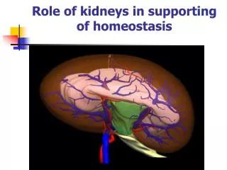
Mechanism of urine forming. Role of kidneys in supporting of homeostasis.
The Nephron Is the Functional Unit of the Kidney • Each kidney in the human contains about 1 million nephrons, each capable of forming urine. The kidney cannot regenerate new nephrons. Therefore, with renal injury, disease, or normal aging, there is a gradual decrease in nephron number.
Each nephron contains (1) a tuft of glomerular capillaries called the glomerulus, through which large amounts of fluid are filtered from the blood, and (2) a long tubule in which the filtered fluid is converted into urine on its way to the pelvis of the kidney.
The macula densa plays an important role in controlling nephron function. Beyond the macula densa, fluid enters the distal tubule that, like the proximal tubule, lies in the renal cortex.
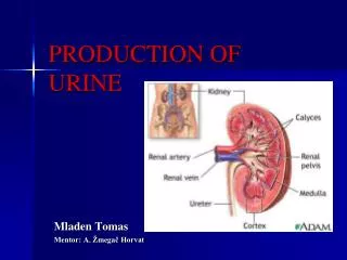
Renal Blood Supply • Blood flow to the two kidneys is normally about 22 per cent of the cardiac output, or 1100 ml/min. • The renal artery enters the kidney through the hilum and then branches progressively to form the interlobar arteries, arcuate arteries, interlobular arteries (also called radial arteries), and afferent arterioles, which lead to the glomerular capillaries, where large amounts of fluid and solutes (except the plasma proteins) are filtered to begin urine formation. • The distal ends of the capillaries of each glomerulus coalesce to form the efferent arteriole, which leads to a second capillary network. the peritubular capillaries, that surrounds the renal tubules. • Video
PHYSIOLOGIC CONTROL OF GLOMERULAR FILTRATION AND RENAL BLOOD FLOW • The determinants of GFR that are most variable and subject to physiologic control include the glomerular hydrostatic pressure and the glomerular capillary colloid osmotic pressure. • These variables, in turn, are influenced by the sympathetic nervous system, hormones and autacoids (vasoactive substances that are released in the kidneys and act locally), and other feedback controls that are intrinsic to the kidneys.
Sympathetic Nervous System Activation Decreases GFR • Strong activation of the renal sympathetic nerves can constrict the renal arterioles and decrease renal blood flow and GFR. • Moderate or mild sympathetic stimulation has little influence on renal blood flow and GFR. For example, reflex activation of the sympathetic nervous system resulting from moderate decreases in pressure at the carotid sinus baroreceptors or cardiopulmonary receptors has little influence on renal blood flow or GFR. Moreover, because the baroreceptors adapt within minutes or hours to sustained changes in arterial pressure, il is unlikely that these reflex mechanisms have an important role in longterm control of renal blood flow and GFR. • The renal sympathetic nerves seem to be most important in reducing GFR during severe, acute disturbances, lasting for a few minutes to a few hours, such as those elicited by the defense reaction, brain ischemia, or severe hemorrhage. In the healthy resting person, there appears to be little sympathetic tone to the kidneys.
Hormonal and Autacoid Control of Renal Circulation • Norepinephrine, Epinephrine, and Endothelin Constrict Renal Blood Vessels and Decrease GFR. Hormones that constrict afferent and efferent arterioles, causing reductions in GFR and renal blood flow, include norepinephrine and epinephrine released from the adrenal medulla. • The endothelin may contribute to hemostasis (minimizing blood loss) when a blood vessel is severed, which damages the endothelium and releases this powerful vasoconstrictor. Plasma endothelin levels also are increased in certain disease states associated with vascular injury, such as toxemia of pregnancy, acute renal failure, and chronic uremia.
Angiotensin II Constricts Efferent Arterioles • A powerful renal vasoconstrictor, angiotensin II, can be considered as a circulating hormone as well as a locally produced autacoid because it is formed in the kidneys as well as in the systemic circulation. Because angiotensin II preferentially constricts efferent arterioles, increased angiotensin II levels raise glomerular hydrostatic pressure while reducing renal blood flow. • It should be kept in mind that increased angiotensin II formation usually occurs in circumstances associated with decreased arterial pressure or volume depletion, which tend to decrease GFR • Increased angiotensin II levels that occur with a low-sodium diet or volume depletion help to preserve GFR and to maintain a normal excretion of metabolic waste products, such as urea and creatinine, that depend on glomerular filtration for their excretion.
Endothelial-Derived Nitric Oxide Decreases Renal Vascular Resistance and Increases GFR • A basal level of nitric oxide production appears to be important for preventing excessive vasoconstriction of the kidneys and allowing them to excrete normal amounts of sodium and water. • Administration of drugs that inhibit the formation of nitric oxide increases renal vascular resistance and decreases GFR and urinary sodium excretion, eventually causing high blood pressure. • In some hypertensive patients, impaired nitric oxide production may contribute to renal vasoconstriction and increased blood pressure.
Prostaglandins and Bradykinin Tend to Increase GFR • Hormones and autacoids that cause vasodilation and increased renal blood flow and GFR include the prostaglandins (PGE 2 and PG12) and bradykinin. • By opposing vasoconstriction of afferent arterioles, the prostaglandins may help to prevent excessive reductions in GFR and renal blood flow. • Under stressful conditions, such as volume depletion or after surgery, the administration of nonsteroidal anti-inflammatory agents, such as aspirin, that inhibit prostaglandin synthesis may cause significant reductions in GFR.
AUTOREGULATION OF GFR AND RENAL BLOOD FLOW • Feedback mechanisms intrinsic to the kidneys normally keep the renal blood flow and GFR relatively constant, despite marked changes in arterial blood pressure. These mechanisms still function in blood-perfused kidneys thal have been removed from the body, independent of systemic influences. This relative constancy of GFR and renal blood flow is referred to as autoregulation. • The primary function of blood flow autoregulation in most other tissues besides the kidneys is to maintain delivery of oxygen and nutrients to the tissues at a normal level and to remove the waste products of metabolism, despite changes in the arterial pressure. In the kidneys, the normal blood flow is much higher than required for these functions. The major function of autoregulation in the kidneys is to maintain a relatively constant GFR and to allow precise control of renal excretion of water and solutes. The GFR normally remains autoregulated (that is, remains relatively constant), despite considerable arterial pressure fluctuations that occur during a person's usual activities. In general, renal blood flow is autoregulated in parallel with GFR, but GFR is more efficiently autoregulated under certain conditions.
Myogenic Autoregulation of Renal Blood Flow and GFR • A second mechanism that contributes to the maintenance of a relatively constant renal blood flow and GFR is the ability of individual blood vessels to resist stretching during increased arterial pressure, a phenomenon referred to as the myogenic mechanism. • Stretch of the vascular wall allows increased movement of calcium ions from the extracellular fluid into the cells, causing them to contract through the mechanisms. This contraction prevents overdistention of the vessel and at the same time, by raising vascular resistance, helps to prevent excessive increases in renal blood flow and GFR when arterial pressure increases.
URINE FORMATION • The rates at which different substances are excreted in the urine represent the sum of three renal processes, (1) glomerular filtration, (2) reabsorption of substances from the renal tubules into the blood, and (3) secretion of substances from the blood into the renal tubules. • Expressed mathematically, • Urinary excretion rate = Filtration rate • - Reabsorption rate + Secretion rate
Urine formation begins with filtration from the glomerular capillaries into Bowman's capsule of a large amount of fluid that is virtually free of protein. • Most substances in the plasma, except for proteins, are freely filtered so that their concentrations in the glomerular filtrate in Bowman's capsule are almost the same as in the plasma.
Why Are Large Amounts of Solutes Filtered and Then Reabsorbed by the Kidneys? • One advantage of a high GFR is that it allows the kidneys to rapidly remove waste products from the body that depend primarily on glomerular filtration for their excretion. Most waste products are poorly reabsorbed by the tubules and, therefore, depend on a high GFR for effective removal from the body. • A second advantage of a high GFR is that it allows all the body fluids to be filtered and processed by the kidney many times each day. Because the entire plasma volume is only about 3 liters, whereas the GFR is about 180 L/day, the entire plasma can be filtered and processed about 60 times each day. This high GFR allows the kidneys to precisely and rapidly control the volume and composition of the body fluids.
Glomerular Capillary Membrane • The glomerular capillary membrane is similar to that of other capillaries, except that it has three (instead of the usual two) major layers: • (1) the endothelium of the capillary, • (2) a basement membrane, and • (3) a layer of epithelial cells (podocytes) surrounding the outer surface of the capillary basement membrane. • Together, these layers make up the filtration barrier that, despite the three layers, filters several hundred times as much water and solutes as the usual capillary membrane.
Glomerular Capillary Membrane • Although the fenestrations are relatively large, endothelial cells are richly endowed with fixed negative charges that hinder the passage of plasma proteins. • The basement membrane effectively prevents filtration of plasma proteins.
The final part of the glomerular membrane is a layer of epithelial cells (podocytes) that encircle the outer surface of the capillaries. The foot processes are separated by gaps called slit pores through which the glomemlar filtrate moves. The epithelial cells, which also have negative charges, provide additional restriction to filtration of plasma proteins. Podocytes
Three basic renal processes • The substance is freely filtered but is also partly reabsorbed from the tubules back into the blood. • For each substance in the plasma, a particular combination of filtration, reabsorption, and secretion occurs. The rate at which the substance is excreted in the urine depends on the relative rates of these three basic renal processes.
Filtration, Reabsorption, and Secretion of Different Substances • In general, tubular, reabsorption is quantitatively more important than tubular secretion in the formation of urine, but secretion plays an important role in determining the amounts of potassium and hydrogen ions and a few other substances that are excreted in the urine. • Most substances that must be cleared from the blood, especially the end products of metabolism such as urea, creatinine, uric acid, and urates, are poorly reabsorbed and are, therefore, excreted in large amounts in the urine. • Certain foreign substances and drugs are also poorly reabsorbed but, in addition, are secreted from the blood into the tubules, so that their excretion rates are high.
Filtration, Reabsorption, and Secretion of Different Substances • Nutritional substances, such as amino acids and glucose, are completely reabsorbed from the tubules and do not appear in the urine even though large amounts are filtered by the glomerular capillaries. Each of the processes - glomerular filtration, tubular reabsorption, and tubular secretion - is regulated according to the needs of the body.
MULTIPLE FUNCTIONS OF THE KIDNEYS IN HOMEOSTASIS • Excretion of metabolic waste products and foreign chemicals • Regulation of water and electrolyte balances • Regulation of body fluid osmolality and electrolyte concentrations • Regulation of acid-base balance • Regulation of arterial pressure • Secretion, metabolism, and excretion of hormones • Gluconeogenesis
Excretion of Metabolic Waste Products, Foreign Chemicals, Drugs, and Hormone Metabolites • The kidneys are the primary means for eliminating waste products of metabolism that are no longer needed by the body. These products include urea (from the metabolism of amino acids), creatinine (from muscle creatine), uric acid (from nucleic acids), the end products of hemoglobin breakdown (such as bilirubin), and metabolites of various hormones. • These waste products must be eliminated from the body as rapidly as they are produced. The kidneys also eliminate most toxins and other foreign substances that are either produced by the body or ingested, such as pesticides, drugs, and food additives.
Regulation of Water and Electrolyte Balances • For maintenance of homeostasis, excretion of water and electrolytes must precisely match intake. If intake exceeds excretion, the amount of that substance in the body will increase. If intake is less than excretion, the amount of that substance in the body will decrease. Intakes of water and many electrolytes usually are governed mainly by a person's eating and drinking habits, necessitating that the kidneys adjust their excretion rates to match intakes of the various substances. • Within 2 to 3 days after raising sodium intake, renal excretion also increases to about 300 mEq/day, so that a balance between intake and output is re-established. However, during the 2 to 3 days of renal adaptation to the high sodium intake, there is a modest accumulation of sodium that raises extracellular fluid volume slightly and triggers hormonal changes and other compensatory responses that signal the kidneys to increase their sodium excretion.
In addition, the kidneys contribute to short-term arterial pressure regulation by secreting vasoactive factors or substances, such as renin, that lead to formation of vasoactive products (for example, angiotensin II). The kidneys play a dominant role in longterm regulation of arterial pressure by excreting variable amounts of sodium and water. Regulation of Arterial Pressure
Regulation of Acid-Base Balance • The kidneys contribute to acid-base regulation, along with the lungs and body fluid buffers, by excreting acids and by regulating the body fluid buffer stores. The kidneys are the only means for eliminating from the body certain types of acids generated by metabolism of proteins, such as sup furic acid and phosphoric acid. kidneys is hypoxia. In the normal person, the kidneys account for almost all the erythropoietin secreted into the circulation. • In people with severe kidney disease or who have had their kidneys removed and have been placed on hemodialysis, severe anemia develops as a result of decreased erythropoietin production.
Regulation of 1,25-Dihydroxy Vitamin D 3 Production • The kidneys produce the active form of vitamin D, 1,25-dihydroxy vitamin D3 (calcitriol) by hydroxylating this vitamin at the number "1" position. • Calcitriol is essential for normal calcium deposition in bone and calcium reabsorption by the gastrointestinal tract. Calcitriol plays an important role in calcium and phosphate regulation.
Glucose Synthesis • The kidneys synthesize glucose from amino acids and other precursors during prolonged fasting, a process referred to as gluconeogenesis. The kidneys' capacity to add glucose to the blood during prolonged periods of fasting rivals that of the liver. • With chronic kidney disease or acute failure of the kidneys, these homeostatic functions are disrupted, and severe abnormalities of body fluid volumes and composition rapidly occur. With complete renal failure, enough accumulation in the body of potassium, acids, fluid, and other substances occurs within a few days to cause death, unless clinical interventions such as hemodialysis are initiated to restore, at least partially, the body fluid and electrolyte balances.
BASIC PRINCIPLES OF OSMOSIS AND OSMOTIC PRESSURE • Osmosis is' the net diffusion of water across a selectively permeable membrane from a region of high water concentration to one thai has a lower water concentration. When a solute is added to pure water, this reduces the concentration of water in the mixture. • If a solute such as sodium chloride is added to the extracellular fluid, water rapidly diffuses from the cells through the cell membranes into the extracellular fluid until the water concentration on both sides of the membrane becomes equal. Conversely, if a solute such as sodium chloride is removed from the extracellular fluid, thereby raising the water concentration, water diffuses from the extracellular fluid through the cell membranes and into the cells. The rate of diffusion of water is called the rate of osmosis.
Isosmotic, Hyperosmotic, and Hypo-osmotic Fluids • Solutions with an osmolarity the same as the cell are called isosmotic, regardless of whether the solute can penetrate the cell membrane. • The terms hyperosmotic and hypo-osmotic refer to solutions that have a higher osmolarity or lower osmolarity, respectively, compared with the normal extracellular fluid, without regard for whether the solute permeates the cell membrane. • Highly permeating substances, such as urea, can cause transient shifts in fluid volumes between the intracellular and extracellular fluids, but given enough time, the concentrations of these substances eventually become equal in the two compartments and have little effect on intracellular volume under steady-state conditions. • Fluid usually enters the body through the gut and must be transported by the blood to all tissues before complete osmotic equilibrium can occur. It usually takes about 30 minutes to achieve osmotic equilibrium everywhere in the body after drinking water.
OSMORECEPTOR-ADH FEEDBACK SYSTEM • 1. An increase in extracellular fluid osmolarity causes the special nerve cells called osmoreceptor cells, located in the anterior hypothalamus near the supraoptic nuclei, to shrink. • 2. Shrinkage of the osmoreceptor cells causes them to fire, sending nerve signals to additional nerve cells in the supraoptic nuclei, which then relay these signals down the stalk of the pituitary gland to the posterior pituitary. • 3. These action potentials conducted to the posterior pituitary stimulate the release of ADH, which is stored in secretory granules (or vesicles) in the nerve endings. • 4. ADH enters the blood stream and is transported to the kidneys, where it increases the water permeability of the late distal tubules, cortical collecting tubules, and inner medullary collecting ducts. • 5. The increased water permeability in the distal nephron segments causes increased water reabsorption and excretion of a small volume of concentrated urine.
ADH Synthesis in Supraoptic and Paraventricular Nuclei of the Hypothalamus and ADH Release from the Posterior Pituitary • The hypothalamus contains two types of magnocellular (large) neurons that synthesize ADH in the supraoptic and paraventricular nuclei of the hypothalamus, about five sixths in the supraoptic nuclei and about one sixth in the paraventricular nuclei. Both of these nuclei have axonal extensions to the posterior pituitary. • Once ADH is synthesized, it is transported down the axons of the neurons to their tips, terminating in the posterior pituitary gland. When the supraoptic and paraventricular nuclei are stimulated by increased osmolarity or other factors, nerve impulses pass down these nerve endings, changing their membrane permeability and increasing calcium entry. ADH stored in the secretory granules (also called vesicles) of the nerve endings is released in response to increased calcium entry. The released ADH is then carried away in the capillary blood of the posterior pituitary into the systemic circulation. Secretion of ADH in response to an osmotic stimulus is rapid, so that plasma ADH levels can increase severalfold within minutes, thereby providing a rapid means for altering renal excretion of water.
A second neuronal area • A second neuronal area important in controlling osmolarity and ADH secretion is located along the anteroventral region of the third ventricle, called the AI/3V region. • Lesions of the AV3V region cause multiple deficits in the control of ADH secretion, thirst, sodium appetite, and blood pressure. Electrical stimulation of this region or stimulation by angiotensin II can alter ADH secretion, thirst, and sodium appetite.
ROLE OF THIRST IN CONTROLLING EXTRACELLULAR FLUID OSMOLARITY AND SODIUM CONCENTRATION • The kidneys minimize fluid loss during water deficits through the osmoreceptor-ADH feedback system. Adequate fluid intake, however, is necessary to counterbalance whatever fluid loss does occur through sweating and breathing and through the gastrointestinal tract. • Fluid intake is regulated by the thirst mechanism, which, together with the osmoreceptor-ADH mechanism, maintains precise control of extracellular fluid osmolarity and sodium concentration. Many of the same factors that stimulate ADH secretion also increase thirst, which is defined as the conscious desire for water.
Central Nervous System Centers for Thirst • Located anterolaterally in the preoptic nucleus is another small area that, when stimulated electrically, causes immediate drinking that continues as long as the stimulation lasts. All these areas together are called the thirst center. • The neurons of the thirst center respond to injections of hypertonic salt solutions by stimulating drinking behavior. These cells almost certainly function as osmoreceptors to activate the thirst mechanism, in the same way that the osmoreceptors stimulate ADH release. • Increased osmolarity of the cerebrospinal fluid in the third ventricle has essentially the same effect to promote drinking. It is likely that the organum vasculosum of the lamina terminalis, which lies immediately beneath the ventricular surface at the inferior end of the AV3V region, is intimately involved in mediating this response.
Stimuli for Thirst • One of the most important is increased extracellular fluid osmolarity, which causes intracellular dehydration in the thirst centers, thereby stimulating the sensation of thirst. The value of this response is obvious: it helps to dilute extracellular fluids and returns osmolarity toward normal. • Decreases in extracellular fluid volume and arterial pressure also stimulate thirst by a pathway that is independent of the one stimulated by increased plasma osmolarity. Thus, blood volume loss by hemorrhage stimulates thirst even though there might be no change in plasma osmolarity. • This probably occurs because of neutral input from cardiopulmonary and systemic arterial baroreceptors in the circulation. A third important stimulus for thirst is angiotensin II. Studies in animals have shown that angiotensin II acts on the subfornical organ and on the organum vasculosum of the lamina terminalis.
Stimuli for Thirst • These regions are outside the blood-brain barrier, and peptides such as angiotensin II diffuse into the tissues. Because angiotensin II is also stimulated by factors associated with hypovolemia and low blood pressure, its effect on thirst helps to restore blood volume and blood pressure toward normal, along with the other actions of angiotensin II on the kidneys to decrease fluid excretion. • Dryness of the mouth and mucous membranes of the esophagus can elicit the sensation of thirst. As a result, a thirsty person may receive relief from thirst almost immediately after drinking water, even though the water has not been absorbed from the gastrointestinal tract and has not yet had an effect on extracellular fluid osmolarity. Gastrointestinal and pharyngeal stimuli influence thirst. For example, in animals that have an esophageal opening to the exterior so that water is never absorbed into the blood, partial relief of thirst occurs after drinking, although the relief is only temporary.
Threshold for Osmolar Stimulus of Drinking • The kidneys must continually excrete at least some fluid, even in a dehydrated person, to rid the body of excess solutes that are ingested or produced by metabolism. Water is also lost by evaporation from the lungs and the gastrointestinal tract and by evaporation and sweating from the skin. Therefore, there is always a tendency for dehydration, with resultant increased extracellular fluid sodium concentration and osmolarity. • When the sodium concentration increases only about 2 mEq/L above normal, the thirst mechanism is activated, causing a desire to drink water. This is called the threshold for drinking. Thus, even small increases in plasma osmolarity are normally followed by water intake, which restores extracellular fluid osmolarity and volume toward normal. In this way, the extracellular fluid osmolarity and sodium concentration are precisely controlled.