Download
1 / 1
20 likes | 441 Views
PMX 8098. Gene expression changes (>2) in HCT116 cells in response to PMX 290. ER Tracker. 4 3 2 1 0 -1 -2 -3 -4. Change in expression (LOG2). Merge. DDIT3 TRIB3 ASNS C20orf139 UPP1 DNAJB9 FGF19 DDIT4 SQSTM1 ATF3 GDF15 BIRC3 SLC7A11 MAP1LC3 PRO618 PPP1R15A KIAA1718

E N D
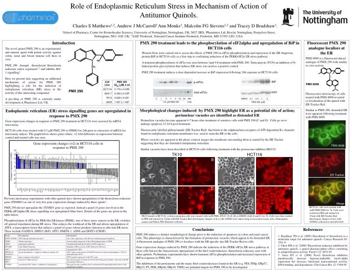
PMX 8098 Gene expression changes (>2) in HCT116 cells in response to PMX 290 ERTracker 4 3 2 1 0 -1 -2 -3 -4 Change in expression (LOG2) Merge DDIT3 TRIB3 ASNSC20orf139 UPP1 DNAJB9 FGF19 DDIT4 SQSTM1 ATF3 GDF15 BIRC3 SLC7A11 MAP1LC3 PRO618 PPP1R15A KIAA1718 SPP1 EGR1 PER1 CCNG2 BHLHB2 TRIM16 TSC22D3 NDRG1 DUSP5 CARD14 HMOx1 BTG1 STC2 DAF GCLM PABPC5 CDKN1A NBR1 RAG1 BLVRB MHC2TA PTGS2 TNFRSF1 HSPA5 MAP2 ATP10B CYP4F2 CLCN3 ILRAP MAT2A NF1 NHLH2 USP44 3-Sep BENE GPR97 CPOX TUBB MTAP SFRS2 GCSH LOC51036 CYP24A1 KITLG POP1 TNFSF18 FABP5 DKK1 Role of Endoplasmic Reticulum Stress in Mechanism of Action of Antitumor Quinols. Charles S Matthews1,2, Andrew J McCarroll2 Ann Monks3, Malcolm FG Stevens1,2 and Tracey D Bradshaw1. 1School of Pharmacy, Centre for Biomolecular Sciences, University of Nottingham, Nottingham, UK, NG7 2RD; 2Pharminox Ltd, Biocity Nottingham, Pennyfoot Street, Nottingham, NG1 1GF, UK; 3 SAIC Frederick, National Cancer Institute-Frederick, Frederick, MD 21702-1201, USA. Introduction PMX 290 treatment leads to the phosphorylation of eIF2alpha and upregulation of BiP in HCT116 cells Fluorescent PMX 290 analogue localises at the ER PMX 8098 is a fluorescent dansyl analogue of PMX 290 with similar in vitro activity. Fluorescence microscopy of cells treated with PMX 8098 revealed co-localisation of the quinol with ER-Tracker Red. Similar to PMX 290, distended ER were apparent following treatment with PMX 8098. The novel quinol PMX 290 is an experimental anti-tumour agent with potent activity against colon, renal and breast tumour cell lines in vitro. PMX 290 disrupts thioredoxin/ thioredoxin reductase redox regulation1,2 and inhibits Hif-1 signalling3. Here we present data suggesting an additional mechanism of action for PMX 290 highlighting a role for the induction of endoplasmic reticulum (ER) stress in the activity of this interesting compound. A pro-drug of PMX 290 is currently under development at Pharminox Ltd, UK. Western blots were carried out to assess the effects of PMX 290 on eIF2a phosphorylation and expression of the ER chaperone protein BiP in HCT116 cells as a first step to confirming induction of the PERK/eIF2a ERstress pathway. A transient phosphorylation of eIF2a was seen between 1and 9 h treatment with PMX 290. Tunicamycin (TUN) an inhibitor of N-linked protein glycosylation that induces ER stress was used as a positive control. PMX 290 treatment induces a dose dependent increase in BiP expression following 24h exposure in HCT116 cells p-eIF2a BiP total eIF2a actin PMX 8098 0 1 5 7 9 16 24 24h 0.5 mM PMX 290 TUN DMSO 0.1 0.5 1 5 PMX 290 (mM) PMX 290 Morphological changes induced by PMX 290 highlight ER as a potential site of action; perinuclear vacuoles are identified as distended ER Endoplasmic reticulum (ER) stress signalling genes are upregulated in response to PMX 290 Gene expression changes in response to PMX 290 treatment in HCT116 were assessed by mRNA microarray. HCT116 cells were treated with 0.5 mM PMX 290 or DMSO for 24h prior to extraction of mRNA for microarray anlysis. The graph below shows genes where >2 fold difference in expression between control and treated cells was seen. Perinuclear vacuoles become apparent 4-7 hours after treatment of sensitive cells with PMX 290 (C and G). Cells go on to undergo apoptosis 12-16 h post-treatment. Fluorescently labelled glibenclamide (ER-Tracker Red) that binds to the sulphonylurea receptors of ATP-dependent K+ channels found in endoplasmic reticulum membranes was used to stain the ER of the cells. Where vacuoles are apparent in the phase contrast images the membrane surrounding them is stained by the ER-Tracker suggesting that they are distended endoplasmic reticulum. Similar vacuoles have been described in HCT116 cells following treatment with the proteasome inhibitor MG132 TK10 HCT116 A B E F C D G H Previous microarray experiments with other quinols have shown upregulation of the thioredoxin reductase gene (TXNRD1) as one of very few gene expression changes induced by these agents1. PMX 290 did not upregulate the TXNRD1 gene as expected. Instead a panel of genes involved in the PERK-eIF2alpha ER stress signalling was upregulated (blue bars). Details of the genes are given in the table below. Phosphorylation of eIF2 by PKR-like ER kinase (PERK), one of three stress sensors in the ER, switches off general translation during ER stress. This reduces the workload of the ER and allows upregulation of ATF4, a transcription factor that induces a panel of genes whose products function to alleviate ER stress. These include GADD34, HSPA5 (BiP), ATF3, HMOX-1, ASNS and DDIT3 (CHOP). HCT116 cells were treated with 1mM PMX 8098 for 7h. Cells were washed in PBS and stained for 15min with ER-Tracker Red (Invitrogen). Images of live cells (1000X) collected as described left. TK10 (renal) or HCT116 (colon) carcinoma cells were treated with 1mM PMX 290 (C, D, G, H) or DMSO (A,B, E and F) for 7h. Cells were then washed in PBS and stained for 15min with ER-Tracker Red (Invitrogen). Images of live cells (400X) were taken using a Leica microscope with a Hamamatsu camera and Nikon NIS-Elements software. Conclusions References 1. Bradshaw TD et al. (2005) Elucidation of thioredoxin as a molecular target for antitumor quinols. Cancer Research 65: 3911-9 2. Chew EH et al. (2008) Thioredoxin reductase inhibition by antitumor quinols: a quinol pharmacophore effect correlating to antiproliferative activity. Faseb J 22: 2072-83 3. Jones DT et al. (2006) Novel thioredoxin inhibitors paradoxically increase hypoxia-inducible factor-alpha expression but decrease functional transcriptional activity, DNA binding, and degradation. Clin Cancer Res 12: 5384-94 PMX 290 induces a distinct morphological change prior to the induction of apoptosis in colon and renal cancer cells. This phenotype is characterised by the formation of perinuclear vacuoles which appear to be distended ER. A fluorescent analogue of PMX 290 co-localises with the ER specific dye ER-Tracker Red in cells. Gene expression changes induced by PMX 290 indicate the induction of the PERK-eIF2a ER stress pathway in these cells but not the characteristic upregulation of the thiol oxidoreductase; thioredoxin reductase seen with other quinols. Preliminary experiments have shown transient eIF2a phosphorylation and increased expression of BiP in response to PMX 290. The inhibition of the proteasome and the many thiol oxidoreductases found in the ER (e.g. PDI, PDIp, ERp57, ERp72, P5, PDIr, ERp46, ERp18, TMX) are potential targets for PMX 290 to be investigated.