Download
1 / 25
500 likes | 973 Views
Dr Jamila EL Medany. ANATOMY OF KIDNEYS. By the end of this course you should be able to discuss : COMPONENTS OF THE URINARY SYSTEM. KIDNEY: SHAPE & POSITION. SURFACE ANATOMY. EXTERNAL FEATURES. HILUM and its CONTENTS. RELATIONS. INTERNAL STRUCTURE. BLOOD SUPPLY

E N D
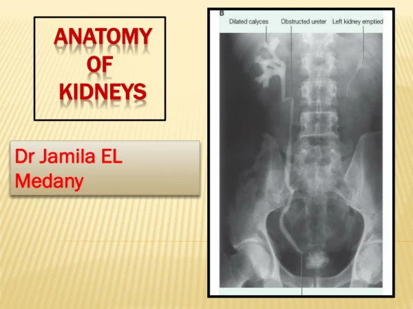
DrJamila EL Medany ANATOMY OF KIDNEYS
By the end of this course you should be able to discuss : COMPONENTS OF THE URINARY SYSTEM. KIDNEY: SHAPE & POSITION. SURFACE ANATOMY. EXTERNAL FEATURES. HILUM and its CONTENTS. RELATIONS. INTERNAL STRUCTURE. BLOOD SUPPLY LYMPH DRAINAGE.. NERVE SUPPLY. Objectives
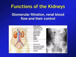
INTRODUCTION • Every day, each kidney filters liters of fluid from the bloodstream. • Although the lungs and the skin also play roles in excretion, the kidneys bear the major responsibility for eliminating nitrogenous (nitrogen-containing) wastes, toxins, and drugs from the body.
Functions: Excretes most of the waste products of metabolism. Controls water & electrolyte balance of the body. Maintain acid-base balance of the blood. Erythropoietinhormone stimulates bone marrow for RBCs formation. Renninenzyme regulates the blood pressure. Converts vitamin D to its active form. Kidney
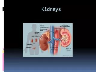
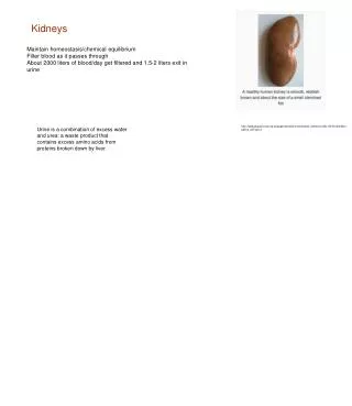
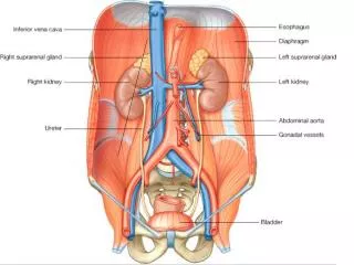
Kidneys are reddish brown in color. Lie behind the peritoneum on the posterior abdominal wall on either side of the vertebral column. They are largely under cover of the costal margin. The right kidney lies slightly lower than the left due to the large size of the right lobe of the liver. The upper border of the right kidney is at the level of 11th intercostal space. The upper border of the left kidney is at the level of 11th rib Kidney
With contraction of the diaphragm the kidney moves downward as much as 2.5 cm. The lateral border is convex, while the medial border is convex at both ends but its middle pat shows a vertical slit called the hilum. The hilum extends into a large cavity called the renal sinus. The hilum transmits the renal vein, two branches of renal artery, ureter, and the third branch of renal artery from the front backward (V.A.U.A.) Kidneys
1- Fibrous capsule: It surrounds the kidney. 2-Perirenal (perinephric) fat : It covers the fibrous capsule 3-Renal fascia: It encloses the kidneys and suprarenal glands. 4-Pararenal (paranephric) fat: It lies external to the renal fascia, and forms part of the retroperitoneal fat. N.B. The last 3 structures support the kidney in position. Coverings
Each kidney has an outer cortex and an inner medulla. Medulla is composed of about 12 renal pyramids. The base of each pyramid is directed toward the cortex & its apex (the renal papilla) is projecting medially. The cortex extends into the medulla between adjacent pyramids as the renal column. Renal Structure
Extending from the bases of the renal pyramids into the cortex are striations known as medullary rays. The renal sinus within the hilum, contains the upper expanded end of the ureter, the renal pelvis. Renal pelvis divides into two or three major calyces, which divides into two or three minor calyces. Renal Structure
Posterior relations • Twelfth rib, • Costodiaphragmatic pleural recess.
4Muscles: Diaphragm Psoas major m., Quadratuslamborum m., Transversusabdominis m. Quadratus lumborum
Posterior Relation 3 Nerves: Subcostal nerve (T12), Iliohypogastric (L1) nerve. Ilioinguinal (L1) nerve
A N T E R I O R R E L A T I O N Right Kidney : • 1- Right suprarenal gland • 2- Liver, • 3- Second partof the duodenum • 4- Right colic flexure • 5- Coils of small intestine Left Kidney : 1- Left suprarenal gland, 2- Stomach, 3- Spleen, 4- Pancreas, 5- Left colic flexure, 6- Descending colon 7- Coils of jejunum
It is the structural and functional unit of the kidney. There are over one million of nephrons in each kidney. Most nephrons are located within the cortex ( cortical nephrons). NEPHRON
Components Of NEPHRON • (a) Glomerulus : • A Knot of capillaries. • (b) Renal Tubule, • Which is composed of : • 1. Glomerular (Bowman’s)) Capsule: • The closed end of the tubule. • It is cup shaped and completely surrounding the glomerulus.
2. Proximal Convoluted Tubule. • 3. Loop of Henle. • 4. Distal Convoluted Tubule. • Collecting (tubules) Ducts: • Each of which receives urine from many nephrons, through the medullary pyramids into the calyces and renal pelvis.
The renal artery arises from the aorta at the level of the second lumbar vertebra. Each renal artery divides into five segmental arteries that enter the hilum of the kidney, four in front and one behind the renal pelvis They are distributed to different segments of the kidney. Lobar artery arises from each segmental artery, one for each renal pyramid. BloodSupply
Each lobar artery gives off 2 or 3 interlobar arteries. The interlobar arteries run toward the cortex on each side of the renal pyramid. Interlobar arteries give off the arcuate arteries at the junction of the cortex and medulla The arcuate arteries give off several interlobular arteries Blood Supply
Interlobular artery gives off afferent glomerular arterioles .
Each nephron is associated with two capillary beds: • Theglomerulus and • Theperitubular capillary bed. • The glomerulus is both fed and drained by arterioles. • The afferent arteriole, which arises from an interlobular artery,is the "feeder vessel," and • the efferent arteriole receives blood that has passed through the glomerulus.
Venous Drainage • Renal vein emerges from the hilum in front of the renal artery and drains into the IVC. • The left renal vein is longer than the right renal vein. • The left renal vein receives the left gonadal & the left suprarenal veins.
Lymph • Lymph Drainage: • Lateral aortic lymph nodes around the origin of the renal artery.
Nerve Supply Renal sympathetic plexus. The afferent fibers that travel through the renal plexus enter the spinal cord in the 10th, 11th, and 12th thoracic nerves. Nerve Supply