Download

1 / 18
180 likes | 233 Views
Learn about serological techniques like cell isolation, separation methods, and monoclonal antibody production by Dr. Dalia Galal. Explore techniques for T-cell subset separation and rosette tests.

E N D
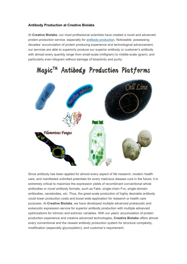
Lecture 10 serology Some different serological techniquesand monoclonal antibody production By dr. Dalia Galal
Serological technique • Isolation of Lymphocyte Populations • Lymphocytes and their specific subpopulations can be isolated by: • Fluorescent activated cell sorter (FACS), • density gradient separation • And rosetting. • Activities for which the lymphocytes can be separated could be: • To detect the ability of a given B cell to produce a given antibody, • To detect the ability of a given T cell to produce particular Cytokines, • To test the ability of a given cell to be stimulated by a given mitogen. • The study of human T cells is best performed using purified cells, since the presence of other cell types may have indirect effects on T cell function.
A. peripheral blood Mononuclear Cells (PBMC) Isolation • The mononuclear cell fraction containing monocytes and lymphocytes is separated from polymorphonuclear cells and red blood cells by density gradient centrifugation. • Counting and markers of cell death Cell suspension (20ul) is diluted with 20ul 0.5% aqueous Trypan blue. • The stained (dead) and non-stained (viable) cells are counted in a haemocytometer.
B. Separation of T and Non - T Cells from Monounclear Cells The E - rosetting Technique The E-rosetting technique describes a procedure for separating T cells and non -T cells from a population of MNCs. • This method is based on the ability of human T cells to bind to sheep erthrocytes via their CD2 molecule. • Neuraminidase treatment of sheep red blood cells (SRBCs) enhances the binding of SRBCs to T lymphocytes: • First neuraminidase treated SRBCs are prepared. • Secondly, SRBCs and MNCs are mixed to form rosettes (E+, which are then isolated from the non - rosetting population (E-, i.e., B cells and monocytes) by Ficoll gradient centrifugation. • In the last step, bound SRBCs are separated from the rosetted T cells by hypotonic lysis (hypotonic buffer capable of lysing red cells, but keeping white cells (or at least nuclei) intact.
C. Separation of T cell subsets • Purification of T - cell populations by indirect antibody panning T cells expressing particular cell surface markers, such as the CD4, CD8, αß - TCR or TCR molecules can be selected by their capacity to bind to an antibody coated plastic plates. • For example to purify CD8+ T cells: • Isolated T cells are treated with a mouse anti - human monoclonal antibody against the CD4 molecule • Then incubated on plastic dishes that have been coated with an anti - mouse IgG antibody. • The T-cell populations that are not CD4 positive (i.e. the αßTCR CD8+ and the γδTCR CD8+ subpopulations), and do not therefore bind the mouse anti human CD4 antibody, will not adhere to the coated plate. • These CD4- cells can be selected physically from the adherent CD4+ subpopulation.
Immunomagnetic Negative Selection of CD4+ T cells • This is another cell separation techniques mediated by antibody-antigen reactions T cells are incubated with specific monoclonal antibodies to surface molecules (anti-CD8) to coat unwanted T cells. • Magnetic beads coated with goat anti-mouse IgG are then applied to the cell suspension in order to bind the antibody coated cells. • After binding; the target cells can be recovered using a strong magnetic field. • Negative isolation is a method by which the CD4+ subset is purified from the CD8* subset binding to the coated magnetic beads. • Furthermore, in a positive selection step, the beads can be removed from the CD8+ target cells by a process of detachment.
Rosette Test Subsets of T lymphocytes • This test can be identified by their differing membrane structures called markers. Markers are categorized as antigen and receptors and can be detected by rosette technique. • The E rosette forming cells were assigned to T cell lineage and the E-rosettes become the principle marker for identification and enumeration of human T cells. • The presence of FC receptors for IgG or IgM on T lymphocyte has been correlated with their functional activity. • Cells with IgM receptors were shown to provide help for B cell differentiation to plasma cell, whereas cells with IgG receptors were reported to function as suppressors. • E-ROSETTE TEST Spontaneous rosette formation with untreated sheep erythrocytes was performed with some modification. • Separate Lymphocytes and adjust the count to 2.5x10⁶ / ml in PBS. • Prepare 1% sheep erythrocyte suspension in PBS after 3 times washing in PBS. • Then 50 micro liters of bovine serum albumin will be taken in tube in which 100µl of lymphocytes suspension and 100ul of 1% sheep RBC suspension will be added. • Then centrifuge for 5 minutes at 1000rpm • After incubation at 40C for 1hour, 0.1% toludine will be added and rosette-forming cells will be counted
Cell sorting in the fluorescence activated cell sorter (FACS). • A suspension of cells is allowed to react with antibodies that are specific for particular molecules on the surface of one of the cell types in the mixture. • The antibody has a fluorochrome attached to it. • The suspension is then mixed with a buffer (sheath fluid) and droplets, each containing a single cell, are generated by ultrasonic vibrations in a nozzle. • The droplets pass one by one through a laser beam, a beam of high intensity light of a particular wavelength. • As the beam hits the cell, two things happen. The fluorochrome molecules absorb the light, but emit light of another wavelength. • The emitted light is focused by collecting lenses on a barrier filter, which only allows light of a certain wavelength to pass through. • Light detectors (photomultipliers) placed behind the barrier filter can then record whether light of a given wavelength has been emitted from the cell and passed through the filter.
At the same time, however, because of the cell curvature and surface unevenness, the light of the laser beam hits the cell at different angles and in turn is reflected from the cell at different angles, i.e. it is scattered. • The character of the light scatter depends on the cell’s size and density; the larger and denser the cell, the more light it scatters. • The degree of light scatter is estimated by measuring light rays reaching the photomultipliers at two different angles in relation to the laser beam; a low angle (a forward scatter) and a right or obtuse angle (side scatter). • The computer then uses these two estimates to determine the size and density of the cell. • Based on this information and information regarding the emission of the fluorescent light, the computer checks whether the cell meets certain criteria for a particular cell type and, depending on the outcome, sends a signal to impart a certain electric charge to the droplet. • As the droplets pass through an electric field generated by the deflection plates, they are sorted according to their charge and collected in tube.
Methods of Monoclonal Antibody Production I- HybridomaTechnique • Large quantities of absolutely pure, specific immunoglobulin directed against an antigen of interest can be produced by fusing a normal plasma cell making the antibody of interest with a myeloma cell with the capacity for prolonged growth in tissue culture. • The resulting mixed cell is called hybridoma. The first stage in making a hybridoma is to generate antibody producing plasma cells. This is done by immunizing a mouse against the antigen of interest and repeating the process several times to ensure that it mounts a good response. • Twoto four days after administration of antigen, the mouse's spleen is removed and broken up to form a cell suspension.
These spleen cells are suspended in culture medium together with a special mouse myeloma cell line. • It is usual to use myeloma cells that do not secrete immunoglobulins since this simplifies purification later on. • Spleen cells are fused with a myeloma cell line by the addition of polyethylene glycol (PEG) which promotes membrane fusion. • Only a small proportion of the cells fuse successfully. The fusion mixture is then set up in culture with medium containing 'HAT'. HAT is a mixture of hypoxanthine, aminopterin and thymidine.
Procedure for making and selecting hybridomas for the production of monoclonal antibodies
II- Recombinant DNA techniques • Attempts are also being made to replace altogether the hybridoma method by recombinant DNA techniques. • One such attempt focuses on the gene segments that specify the fragment antigen binding (Fab) of an immunoglobulin molecule, the VH CH1 and VLCL. • These segments can be amplified by PCR from many different mRNA (cDNA) molecules expressed in a population of cells undergoing an immune response. The amplified segments are inserted into a suitable vector, cloned and paired randomly (always one, VH CH1 with VLCL, in a suitable vector) and the pairs translated into proteins (Fabs).
Screening of this combinatorial library of antibodies with labeled antigen then identifies these combinations that bind this antigen. • The identified VHCH1-VLCL pairs are placed in to an expression vector, either bacterial or mammalian, and used to produce large quantities of antibodies with selected specificity.
Uses of monoclonal antibodies • The greatest impact of Monoclonal antibodies in immunology has been on the analysis of cell membrane antigens. • Because Monoclonal antibodies have a single specificity compared to the range of antibody molecules present in the serum, monoclonal antibodies have multiple clinical applications including: • o Identifying and quantifying hormones • o Typing tissues and blood • o Identifying infectious agents • o Identifying clusters of differentiation for the • classification and follow-up therapy of • leukemias and lymphomas • o Identifying tumor antigens and autoantibodies • o Immunotherapy