THE HEART
240 likes | 562 Views
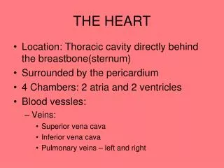
THE HEART. Location: Thoracic cavity directly behind the breastbone(sternum) Surrounded by the pericardium 4 Chambers: 2 atria and 2 ventricles Blood vessles: Veins: Superior vena cava Inferior vena cava Pulmonary veins – left and right. Arteries: Aorta Pulmonary

THE HEART
E N D
Presentation Transcript
THE HEART • Location: Thoracic cavity directly behind the breastbone(sternum) • Surrounded by the pericardium • 4 Chambers: 2 atria and 2 ventricles • Blood vessles: • Veins: • Superior vena cava • Inferior vena cava • Pulmonary veins – left and right
Arteries: • Aorta • Pulmonary • Blood is pumped through the chambers, aided by four heart valves • The valves open and close to let the blood flow in only one direction
Valves: • Atrioventricular valves: • Tricuspid • Bicuspid (mitral) • Semilunar valves • Aortic • Pulmonary • Where • are the four heart valves? • The tricuspid valve is between the right atrium and right ventricle • The pulmonary (semilunar) valve is between the right ventricle and the pulmonary artery
The bicuspid (mitral) valve is between the left atrium and left ventricle. • The aortic valve is between the left ventricle and the aorta. • Valves open when the pressure in the preceding chamber is higher than that in the next chamber and they close when pressure in the preceding chamber falls below that in the next chamber. • What causes the familiar “lubb-dupp” sounds associated with the heart?
The closing of the heart valves causes blood to splash against the valves. This causes the heart sounds. • Each heart beat is one cardiac cycle. • There are three parts to each cycle: • Part 1: two atria contract (systole) at once. two ventricles are relaxed (diastole). • Part 2: two ventricles contract at once. two atria are relaxed
Part three: all chambers relax • The heart contracts and relaxes through this cycle on average about 70 times a minute • Each heart beat lasts just under a minute. • Blood flow during the beat: • Atrial systole – ventricular filling • Ventricular systole – blood forced through semilunar valves into circulation
The Meaning of Blood Pressure • Pressure = force of blood pushing against blood vessel wall • Example: 120/80 • Systolic number = pressure of ventricular contraction • Diastolic number = pressure when heart is relaxed
Intrinsic Control of Heartbeat • Cardiac conduction system • SA node • AV node • AV bundle • Left and right bundle branches • Purkinge fibers • Allows impulse conduction to spread first to atria and then after delay to ventricles • Allows for adequate filling of all chambers
Extrinsic Control of Heartbeat • Medulla oblongata (in the brain stem) • Norepinephrine – released by the adrenal medulla (on top of kidney) during exercise, can increase heart beat
Electrocardiogram • P wave: occurs just before the atria contract • QRS complex: occurs just before ventricular contraction • T wave: ventricular relaxation
Blood Vessels • Arteries • Take blood away from the heart • Thick (three layers) and muscular • Blood enters arteries under pressure • Veins • Channel blood towards the heart • Thinner walls • Capillaries • Walls are only one cell thick • Precapillary sphincters regulate flow into capillary beds
Blood Flow back to the Heart • How does blood get back to the heart with such little pressure? • Valves play a big role • Muscular pumps
Pathway of the Blood • Pulmonary circuit (to lungs) • Systemic circuit (to body tissues) • Coronary arteries supply heart muscle with blood
Pulmonary Circulation • Right atrium – from superior vena cava • Right ventricle • Pulmonary trunk – semilunar valve • Pulmonary arteries • Lungs (pulmonary capillaries) • Pulmonary veins – oxygen rich • Left atrium
Systemic Circulation • Left atrium • Left ventricle • Aorta • System arteries • Systemic capillaries • Systemic veins • Superior vena cava • Right atrium
Cardiovascular disease • #1 killer; 1 in 2 Americans die of it • High blood pressure ( hypertension) • Damages organs, especially heart, kidneys • Contributes to atherosclerosis, stroke risk • Heart disease – atherosclerosis • Narrowing of arteries – lipid buildup, thickening of walls • Coronary arteries especially susceptible • Restricted blood flow angina • Blocked flow heart attack • Treatments: balloon angioplasty, coronary bypass
Heart muscle dies after 2 hrs without oxygen • Signs of heart attack: chest pains, nausea, and dizziness • Stroke = cutoff of oxygen to brain cells – cell survival – only 5 minutes • Taking care of your heart • Exercise (aerobic, at least 20 minutes) • Good nutrition (control fats & body weight) • QUIT SMOKING
Summary of Main Points • The heart is a double pump that operates continuously • Arteries take blood away from the heart • Veins take blood back to the heart • Capillaries are the exchange vessels • Pulmonary and systemic circulations serve the lungs and body respectively • Blood consists of plasma, oxygen-carrying red blood cells, and defensive white blood cells • Cardiovascular disorders can be genetic or a result of poor decisions


