Download
1 / 46
490 likes | 755 Views
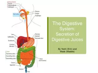
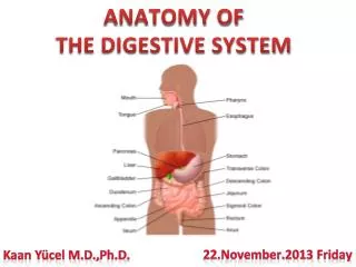
Anatomy of the Digestive System. Anatomy & Physiology Chapter 25. Function. Altering the chemical & physical composition of food so that it can be absorbed & used by body cells (digestion). Mouth Oropharynx Esophagus Stomach Duodenum Jejunum Ileum Large Intestine Cecum Colon

E N D
Anatomy of the Digestive System Anatomy & Physiology Chapter 25
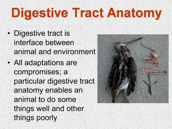
Function • Altering the chemical & physical composition of food so that it can be absorbed & used by body cells (digestion)
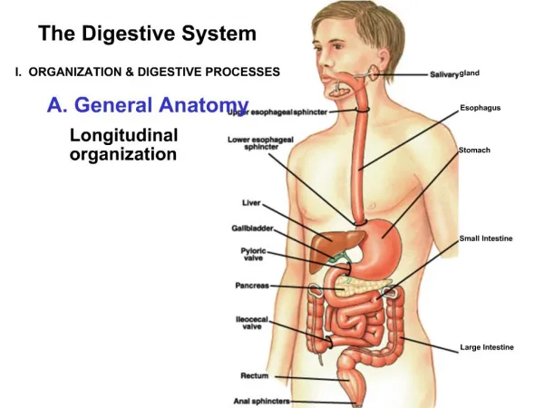
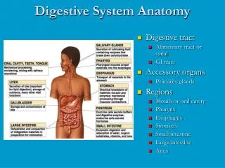
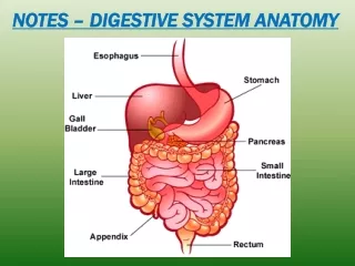
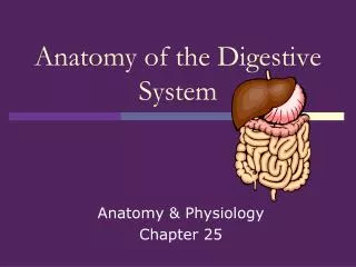
Mouth Oropharynx Esophagus Stomach Duodenum Jejunum Ileum Large Intestine Cecum Colon Ascending colon Transverse colon Descending colon Sigmoid colon Rectum Anal Canal Accessory Organs Salivary glands Parotid Submandibular Sublingual Tongue Teeth Liver Gallbladder Pancreas Vermiform appendix Organs of the Digestive System
Walls of the Gastrointestinal (GI)Tract • Tube with 4 layers of tissue • Mucosa • Submucosa • Muscularis • Serosa
Mucosa • Inner most layer • Made of 3 layers of epithelium, connective tissue & smooth muscle
Submucosa • Thicker than mucosal layer • Connective tissue layer that contains glands, blood vessels, nerve plexus (Meissner plexus)
Muscularis • Thick layer of muscle tissue • Inner layer of circular smooth muscle • Outer layer of longitudinal smooth muscle • Myenteric plexus between the muscular layers
Serosa • Outermost layer • Made of connective tissue & peritoneum (visceral layer) • Layer of peritoneum that lines the abdominal cavity= parietal layer • Mesentery is the fold of membrane that connects the parietal & visceral layer of peritoneum
Mouth (Oral cavity) • Lips • Cheek • Tongue • Hard & Soft Palates
Lips • Surround the orifice of the mouth & form anterior boundary • Covered by skin externally & mucous membrane internally • Philtrum: shallow vertical groove that marks the midline of upper lip
Cheeks • Form lateral boundaries, continuous with lips, lined by mucous membranes • Formed in large part by buccinator muscle
Hard & Soft Palates • Hard palate: consists of 4 bones: 2 maxillae & 2 palatines • Soft palate: partition between mouth & nasopharynx • Uvula: small cone shaped process extending from soft palate
Tongue • Intrinsic muscle: changes in size & shape of tongue; important for mastication (chewing) • Extrinsic muscle: origin outside the tongue; important for deglutition (swallowing) & talking • 3 parts: root, body, tip
Papillae • Vallate: large, form an inverted V on posterior part of tongue; 10-14; taste buds on lateral aspect • Fungiform: taste buds on lateral aspect; mostly on sides & tips of tongue • Filiform: no taste buds, over anterior 2/3 of tongue; whitish appearance
Lingual Frenulum • Fold of mucous membrane on the undersurface of the tongue that anchors the tongue to the floor of mouth
Salivary glands • Three pairs: • Parotid • Submandibular • Sublingual • Secrete about 1 L of saliva/day
Parotid Glands • Largest • Between skin & masseter muscle in front of & below the ear • Produce a serous (watery) type of saliva
Submandibular glands • Mixed gland-contain both serous & mucus-producing elements • Located below mandibular angle
Sublingual glands • Smallest • Under the mucous membrane covering the floor of the mouth • Produce only a mucous type of saliva
Tooth • 3 main parts • Crown: exposed portion, covered by enamel • Neck: area surrounded by gingiva • Root: area that fits into jaw • Tooth suspended in place by periodontal membrane
Tooth structure • Dentin: makes up greatest portion of tooth shell, covered by enamel on crown & cementum in neck & root • Dentin contains a pulpcavity consisting of vessels & nerves
Type of teeth • Deciduous teeth (baby): 20; erupt from 6 mos. to 20 mos. • Permanent teeth: 32
Pharynx • Food now called a bolus leaves mouth & enters oropharynx • Food does not go thru nasopharynx
Esophagus • Collapsible tube, posterior to trachea • Mucosa: stratified squamous epithelium to resist abrasion • Muscularis: striated in upper third, smooth in lower third
Sphincters of esophagus • Upper esophageal sphincter: helps prevent air from entering during respiration • Lower esophageal sphincter (cardiac sphincter): between stomach & esophagus
Stomach • Three divisions: • Fundus: enlarged portion to left & above opening of esophagus • Body & Pylorus
Sphincter of Stomach • Pyloric sphincter: controls opening of pylorus to duodenum
Gastric mucosa • Folds (rugae) with depressions (gastricpits) • Gastric glands are located below the level of the pits
Gastric glands • 3 major secretory cells: • Chief cells: secrete enzymes • Parietal cells: secrete HCl & intrinsic factor (binds to Vitamin B12 to protect it) • Endocrine cells: secrete ghrelin (stimulates hypothalamus to increase appetite) & gastrin (regulates gastric function)
Gastric muscle • Made of 3 layers instead of 2
Functions of stomach • Reservoir • Secretes gastric juice to aid in digestion • Churns food • Secretes intrinsic factor • Absorption-small amounts • Produces hormones • Protects by destroying pathogens
Small intestine • Main site of digestion & absorption • 3 divisions: • Duodenum • Jejunum • Ileum
Wall of Small Intestine • Has circular folds (plicae) with many tiny projections (villi) • Each villus contains vessels & lacteal • Epithelial cells on surface of villi have microvilli which form a brush border which increases surface area
Goblet cells • Large numbers of mucus secreting goblet cells on villi & in crypts • Crypts serve as area of rapid mitotic division & at base of crypts secretory cells produce an enzyme that is thought to inhibit bacterial growth
Large Intestine • Divisions: • Cecum: blind pouch • Colon • Ascending colon: right side, ileum attaches at junction of cecum & ascending colon • Transverse colon: from hepatic flexure to splenic flexure • Descending colon: left side • Sigmoid colon: S shaped • Rectum: terminal inch is anal canal
Wall of Large Intestine • Intestinal mucus glands which produce mucus to lubricate feces • Longitudinal muscle fibers form strips called taeniae coli & circular muscles are grouped into rings that produce pouches, haustra
Vermiform appendix • Wormlike tubular organ, communicates with cecum
Peritoneum • Large continuous sheet of serous membrane that lines the walls of abdominal cavity & forms outer serous coat of organs • Mesentery: fan shaped projection of peritoneum encloses the jejunum & ileum • Greater omentum: continuation of serosa of stomach to transverse colon • Lesser omentum: from liver to lesser curvature of stomach
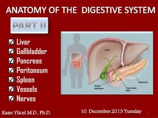
Liver • Largest gland in body • 2 lobes separated by falciform ligament: • Left lobe (about 1/6) • Right lobe
Hepatic lobules • anatomical units of liver • Branch of hepatic vein through center of each lobule • Outer corners of lobules are the branches of hepatic artery, portal vein, hepatic duct
Bile ducts • Small bile ducts join to form right & left hepatic duct which join to form hepaticduct which merges with cystic duct from gallbladder to form common bile duct which opens into duodenum at majorduodenal papilla
Functions of liver • Detoxify substances • Secrete bile • Metabolism of foods • Store several substances • Produces plasma proteins & site of hematopoiesis during fetal development
Gallbladder • Pear shaped on underside of liver • Serous, muscular & mucosal layer • Functions: stores, concentrates & ejects bile
Pancreas • In curve of duodenum, extending behind stomach • Exocrine gland (most) • Acinar cells: secrete enzymes through pancreatic duct that empties into duodenum • Endocrine gland-Islets • Alpha cells: produce glucagon • Beta cells: produce insulin
Image Citations • Slide 6: Brunner’s glands, 2/28/07, http://w3.ouhsc.edu/histology/Text%20Sections/Lower%20GI.html • Slide 25: Lower esophageal sphincter, 3/13/07, http://hopkins-gi.nts.jhu.edu/pages/latin/templates/index.cfm?pg=disease1&organ=1&disease=13&lang_id=1 • Slide 38: The human vermiform appendix, 3/13/07, http://www.talkorigins.org/faqs/vestiges/appendix.html