Download
1 / 15
150 likes | 370 Views
Work-up of Palpable Breast Mass. Courtney Riley Radiology August 28, 2007. Palpable Breast Mass in the Non-Pregnant Female. Examine patient If mass is round, well defined and mobile: <20 yo f/u in 2-6 wks with U/S if persists 20-25yo f/u 2-6 wks with U/S +/- mammogram

E N D
Work-up of Palpable Breast Mass Courtney Riley Radiology August 28, 2007
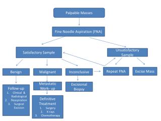
Palpable Breast Mass in the Non-Pregnant Female • Examine patient • If mass is round, well defined and mobile: • <20 yo f/u in 2-6 wks with U/S if persists • 20-25yo f/u 2-6 wks with U/S +/- mammogram • >25yo cautiously f/u 2-6 wks with mammogram +/- U/S Courtesy of Dr. J Fulton
Palpable Breast Mass in Non-Pregnant Female • Hard, Fixed mass: • <20 yo → ultrasound • 20-25 yo → if negative family history and low suspicion cautious f/u in 2-6 weeks; if strong family history and high suspicion U/S +/- mammogram • >25 yo → mammogram + U/S → if cystic w/u complete; if complex cyst or solid mass → biopsy Courtesy of Dr. J Fulton
For cystic mass: Painful → aspirate Nontender → work up complete For complex cyst or solid mass: Biopsy Palpable Breast Mass in Pregnant Patient Ultrasound first Courtesy of Dr. J Fulton
Interpretation of Mammogram ~ Note any asymmetry between breast & density of tissue ~ Calcifications: - Normal with age → round, > 2mm in diameter, or calcifications in vessel walls - Suspicious → fine (<1mm) sandlike, microcalcifications ~ Note signs associated with malignancy: - Focal skin thickening or dimpling, unilateral nipple retraction, vascular asymmetry ~Compare findings to old films Mettler: Essentials of Radiology, 2nd ed.
Non-palpable Mass Noted on Mammogram for Palpable Breast Mass
Spiculated Mass • Differential diagnosis for a spiculated mass visible on mammogram includes malignancy, post-surgical scar, radial scar, hematoma, or past trauma tothe breast.