Download
1 / 32
490 likes | 2.37k Views
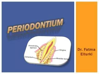
ANATOMY OF PERIODONTIUM PART II. DR.HOUNIDA IDRIS. CEMENTUM. DEFINITION : it is a thin layer of hard mineralized tissue ( bone like tissue) that covers the surface of the tooth. Characteristics: It is lightly yellow and softer than dentine.

E N D
ANATOMY OF PERIODONTIUM PART II DR.HOUNIDA IDRIS
DEFINITION: it is a thin layer of hard mineralized tissue ( bone like tissue) that covers the surface of the tooth.
Characteristics: • It is lightly yellow and softer than dentine. • It overlies and is attached to the dentine of the root. • It’s a bone like tissue that is more resistant to resorption than bone. • Cementum dose not have its own blood or nutrients from the periodontal ligament
Function: • It gives attachment to the collagen fibers ligament , without cementum the toot would fall out of its socket. • The outer layer of cementum protects the underline dentine and seals the ends of the open dentinal tubules. • Cementum formation compensate for tooth wear at the occlusal or incisal surface due to attrition .
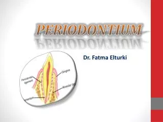
DEFINITION: It is a layer of soft connective tissue that covers the root of the tooth and attaches it to the bone of tooth socket ( alveolar bone). • Consist: it consists of bundles of collagen fibers arranged into a network referred as principle fibers.
Function: • Support and maintains the tooth in its socket. • Provides sensory feeling to the tooth such pressure and pain sensation. • Provide nutrition to the cementum and the alveolar bone of the tooth socket. • It builds and maintains cementum and the alveolar bone of the tooth socket. • Resorptive function: Can remodel to pressure such as that applied during orthodontics treatment (braces).
PDL fibers: • It ranges in thickness. • the width decrease with age. • Tissue with high turnover rate.
Sharpesy’s fibers: • it’s a fibers that insert into the cementum and bone.
DEFINITION: • The alveolar process or the alveolar bone is the bone of the upper and lower jaw that surrounds and support the roots of the teeth.
Characteristics: • The existence of the alveolar process is depend on the teeth ; when the teeth are extracted the alveolar bone resorbes. If the teeth do not erupt the alveolar bone dose not develop. • Alveolar bone is mineralized tissue made by cells called osteoblast and osteolcalst. • The alveolar bone has blood vessels and nerve innervations.
Functions: • It form the bony socket that provides support and protection for the roots of the teeth. • Remodels in response to mechanical forces and inflammation.
Layers : • Cortical plate. • Spongy bon. • Lamina dura : thin compact bone that lines the tooth socket and has many small openings for blood vessels and nerve fibers. On an x-ray a lamina dura will appear as a radiopaque line surrounding the tooth root. An intact lamina dura is seen as a sign of healthy periodontium. Lamina dura, along with the periodontal ligament, plays an important role in bone remodeling and thus in orthodontic tooth movement
Nerve supply to the periodontium: • Is derived from the branch of the trigeminal nerve.
BLOOD SUPPLY TO THE PERIODONTIUM • The vessels of the periodontium anastomes ( join together) to create a complex system blood vessels that supply tissues ( hard and soft). • The major function of the complex network of blood vessels of periodontium is to transport oxygen and nutrients to the tissues cells and remove carbon dioxide and other products from the cells.
Maxilla ( upper jaw): • Maxillary gingiva, periodontal ligaments and alveolar bone are supplied by: • Anterior and posterior superior alveolar arteries. • Infraorbital artery. • Greater palatine artery.
Mandible ( lower jaw ) • Mandibuler gingiva,periodontal ligaments and alveolar bone: • Inferior alveolar artery. • Branches of the inferior alveolar artery: the buccal, facial, mental and sublingual arteries.
Vascular supply to the teeth and periodontal tissues: • Superior alveolar arteries Maxillary periodontal tissues. • Inferior alveolar artery Mandibular periodontal tissues.