Download
1 / 28

E N D
INTRODUCTION Definition: A science: study normal micro-structure & its related function of human body. 4 structural levels:Cell: the smallest structural & functional unit. Tissue:groups ofcells (similar in morphology or related in function)+intercellular materials 4 types of fundamental tissue epithelium connective tissue muscular tissue nervous tissue
Organs:organizations of various kinds of tissues in particular ways & perform a specificfunction. System:formed by several function-related organs which together perform a continuous physiological function. For example: digestive system
Why to study histology ? • To complete the knowledge of human body’s structures----from gross to microscopic • Be able to understand how the different tissues function----the basis of physiology • Can find the diseases only after the normal is known----the basis of pathology • It is related to some modern science fields: cell apoptosis, cell recognition, implantation of embryo stem cells, eugenics and etc. • It is also a foundation of clinic sciences—for a good doctor needed in futrue
Unitused in microscope 1μm=1/1000 mm 1 n m=1/1000μm Maximumresolution Light microscope: 0.2μm Transmission electron microscope: 0.2nm Scanning electron microscope: 5nm
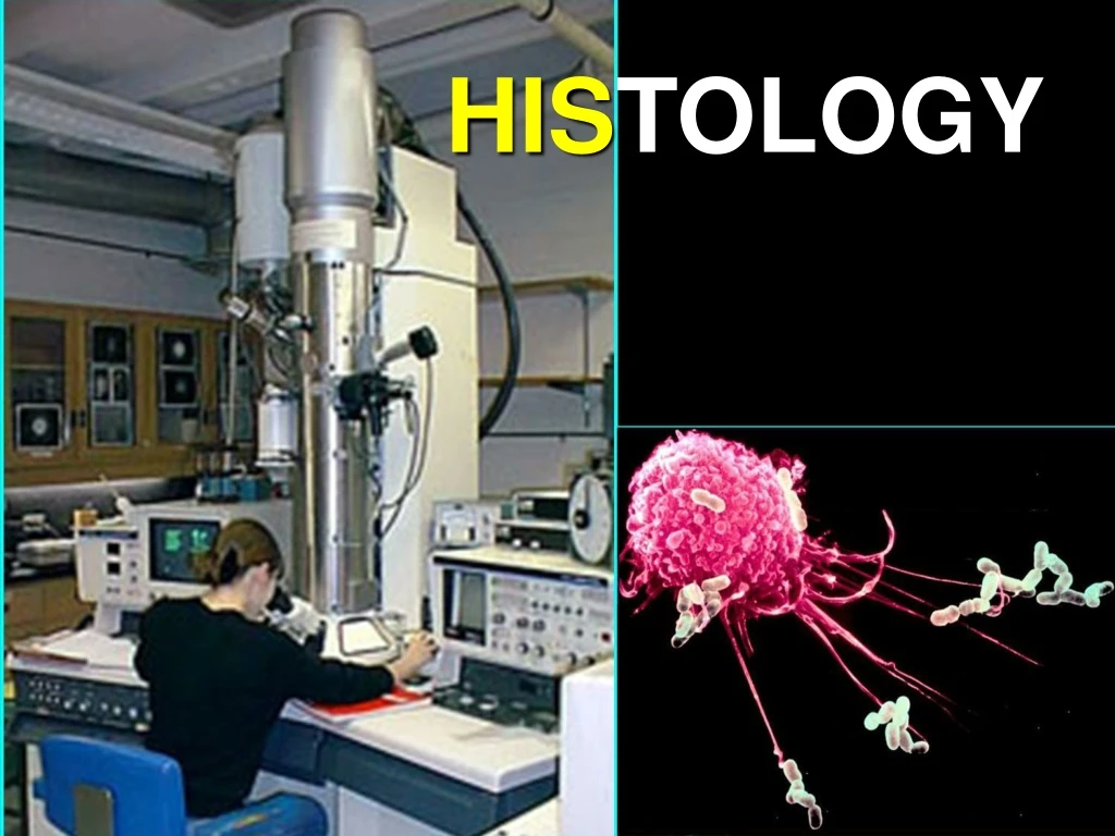
I. Light microscopy Investigative methods of histology
1. Tissue preparation A. paraffin section preparation • Specimen:as fresh as possible • Fixation: fixative: formalin solution; purpose: to preserve the structural organisation • Dehydration: replace the water in the tissue by alcohol • Clearing: replace the alcohol by xylene • Embedding: replace the xylene w/ melted paraffin • Sectioning & mounting l
B.Frozen section: Better for preserving chemical components (e.g. enzymes) Freezing→cryotomy→staining C. the others: Smear preparations: for blood etc; Grind preparations: for bone
2. Staining • Purpose: To make tissue section pigment for observation. • H-EStaining: Hematoxylin:basicdye,purple-blue Eosin: acid dye, pink color Basophilic: components bonded by basic dye (H); pruple-blue(nuclear chromatin & basophilic substance in cytoplasm) Acidophilic: components bonded by acidic dye (E); pink (cytoplasm & collagenous fiber)
Neutrophilic: do not stain w/ both basic and acid dyes Metachromasia: a dye stains tissue a different color from that of dye solution, e.g. toluidine blue stains mast cells in purple color Special staining: Argyrophilia Fluorescent staining
HO (Hoechst 33258)-PI staining shows the apoptosis in human HL-60 cells Silver staining of the neuron and the bile canaliculi
II. Electron Microscopy 1. Transmission Electron Microscope (TEM) • Using a beam of electrons (short wave-lengths) instead of visible light. • Sectionpreparation: similar to those for L.M mainly, plastic instead of paraffin, 50-70nm thick, heavy metal salts instead of HE. • Resolution: 0.1-0.5nm (0.2nm) • Ultrastructrue:The structure in EM • Electron-dense / electron-lucent
2. Scanning Electron Microscope (SEM) Sowing the 3 dimensional surface Architecture of cells and tissues Resolution: 5nm
III.Histochemistry & Cytochemistry Reveal the chemical composition in situ (e.g. proteins, a.a., nucleic acid, lipids, enzymes etc.) w/ chemical, biochemical methods. • The product of chemical reaction should be insoluble / colored / electron- scattering, & be seen in LM or EM
For instance: • PAS(Periodic Acid Schiff) reaction: for manifesting polysaccharide and proteoglycan (e.g. glycogen). polysaccharide + HIO4 (hydroxyl group) (oxidise) Aldehyde group + Shiff’s reagent (colorless) Purplish red depositor
Immunocytochemistry Based on antigen binds to specific antibody. Tissue section w/ Antigen +labelledantibody labelled Ag-Ab complex Fluoresceinlabelling enzyme labelling colloidal goldlabelling
NOS & GnRH positive Neurons in hypothalamus of rat NOS positive neuron in hypothalamus ofrat
IV. tissue culture V. isotopic tracing VI. in situhybridization (ISH) —nucleic acid molecular hybridization Use nucleotide probe to check target fragment of intracellular DNAor mRNA in situ, in order to study the gene expression.
Expression of PSA and PSAmRNA in human prostate (histochemistry & ISH)
Themethod in learninghistology • Combination of the 2 dimentional structure with 3 dimentional
Combination of the theory with practice • Combination of the structure with function • Concern the dynamic change