Download
1 / 35
400 likes | 989 Views
Atrial Septal Defects. Dr Nithin P G. Preview. Introduction Embryology & Types of ASD Physiology, natural history & clinical features Investigations -salient features Management. References. Joseph Perloff . The clinical recognition of congenital heart disease. Fifth Edition 2003

E N D
Atrial Septal Defects Dr Nithin P G
Preview • Introduction • Embryology & Types of ASD • Physiology, natural history & clinical features • Investigations -salient features • Management
References • Joseph Perloff. The clinical recognition of congenital heart disease. Fifth Edition 2003 • Abraham M. Rudolf. Congenital diseases of the heart. Third Edition 2009 • Moss & Adams’ Heart diseases in infants, children & Adolescents. Seventh Edition 2008 • Nadas' Pediatric Cardiology. Second Edition 2006 • ACC/AHA 2008 Guidelines for the Management of Adults With Congenital Heart disease • ESC Guidelines for the management of grown-up congenital heart disease 2010
Introduction • 6-10% of all cardiac anomalies(3) • M:F = 1:2 [ sinus venosus defects 1:1](1)(4) • 1 in 1500 live births(3) • Most common congenital abnormality in adult > 40yrs [30-40%]
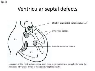
Embryology & types of ASD • Embryology [video] • Types of ASD • Secundum ASD • Primum ASD • Sinus venosus ASD • Coronary Sinus type • PAPVC
Embryology & types of ASD Associated anomalies(5) • 30% cases have associated anomalies • Secundum- Valvular PS, MVP, PAPVC • Primum- Cleft MV, Discrete subaortic AS • SV- PAPVC • Coronary Sinus- PAPVC, PLSVC
Embryology & types of ASD Inherited disorders- secundum ASD(3) • Holt-Oram syndrome [AD] • TBX5 • Familial syndromes • NKX2.5 and GATA4 • missense mutation in myosin heavy chain 6
Physiology, natural history Shunt (2) • Size of defect • As large as mitral valve orifice • Ventricular compliance [ most important]
Physiology, natural history Effects of changes in SV of left and right ventricles associated with changes in PVR and with changes in compliance of the respective ventricles after birth in the presence of an ASD(2)
Physiology, natural history ASD shunt flow during cardiac cycle(3)
Physiology, natural history • Effects of L to R shunt(2) • RA/RV dilation, volume overload RV, RVF • Atrial arrhythmias • Increased PBF • +/- PAH
Physiology, natural history ASD & PAH • Not all individuals develop PAH 702 pts isolated ASDs [Secundum or SV]. 40 (6%) had PVOD, defined as a TPR of >7 U/m2 ….34 women (85%) and 6 men… < 19 years of age no PVOD. Circulation 1987;76:1037-1042 • Why others are spared? • Factors • Increased PBF • Coexistant PAH • Concominant adult heart diseases
Physiology, natural history Clinical course(1) (2) (3) (4) (5) • Infancy • Child hood • Adults
Clinical Findings • G/E- thin, upper limb abnormalities • JVP- mean normal, a=v • Pulse & BP- no change in pulse volume and BP during valsalva, square wave response • Prominent RV pulsations, 2nd space pulsations • Auscultation • Loud T1 • Wide fixed split S2 • Pulmonary ESM • Tricuspid MDM • Features of PAH in some cases
Special situations • Lutembacher- RHD MS/MR + secundum ASD • PAPVC & Scimitar syndrome • Raghib’s syndrome- coronary sinus ASD + PLSVC
Investigations- salient features ECG • SN dysfunctions • AF, AFL, SVT • Absence of sinus arrhythmia • AV conduction • Increased PR interval • CHB • rsR’ or rSr’ pattern in V1 • Crochetage [notched R II,III,aVF]
Investigations- salient features CXR • RAE • RV Apex • Prom MPA • Increased PBF
Investigations- salient features Echo • Most important Ix for confirmation of diagnosis, location and type of ASD, severity, need for intervention & planning further Mx/ Follow up • RA, RV Volume overload • Associated abnormalities • Abnormal venous connections • Suitability for device closure, rims
Investigations- salient features Bi-caval view for IVC & SVC rims [subcoastalsagittal & TEE]
Investigations- salient features AV rim & Postero-superior (atrial) rim [Suboastal coronal & TEE 4C] Aortic rim [TTE PSX & TEE BSX]
Investigations- salient features CATH • Main indication is assessment of PVR in doubtful cases & anomalous venous connections not visible on echo • Oximetry • SVC step up 10% or 5% in 2 serial samples [AVSD, RSOV/LV to RA, VSD + TR, PAPVC, Systemic AVF] • SVC sat >75-80% [RPV to SVC, LPV to L inominate vein] • CS sat > 45% PLSVC or anomalous PV connections to CS • Pressures • RA, LA mean normal, RA a=v • RVSP 35-40 mm Hg in infants & 25-30 mm Hg in Adults, diff of 15-30 mm b/w RVSP & PAP noted
Management • In whom, When & How should you close the defect ? • RV volume overload [+/- symptoms] • Paradoxical embolism • Surgical vs Device closure
Management(5) (6) • <5 mm, no volume overload [unless paradoxical embolism] followed up • Secundum ASD>5 mm, <38 mm size with a rim of 5 mm all around except towards aorta Device closure [Larger ASD, associated tricuspid repair, sinus venosus, coronary sinus, or primum ASD for Sx] • PAP <5 WU, if>5 WU then PVR<2/3 SVR, PAP<2/3 SYS Pressure [baseline or when challenged with vasodilators with net Qp: Qs>1.5] (6)
Occluder diameter 2-4mm size larger than the maximum strechable defect size
Complications Procedural success rate 95%-98% Very rare complication (<0.5%)Rev EspCardiolCardiol. 2003;56:383; (5) (6) • Entrapment in RA structures & PV, impingement of aorta • Inability to release, withdraw • Twisting of device • Dislodgment & embolization • Thrombosis Very rare [0.05-0.2%] J Am CollCardiol 2004;43:302 Related to • Poor implant apposition • Poor device endothelialization • Underlying prothrombotic
Follow up • Issues(5) (6) • Atrial arrhythmias- OAC, RF ablation • RV & LV dysfunction [Pre intervention- balloon occlusion and reassessment of hemodynamics in poor LV function(6)] • PAH • Coexistantvalvular or other cardiac lesions • IE prophylaxis upto 6m after procedure • Aspirin atleast 100mg daily upto 6 m (6) • Echo f/up at 24 hrs, 1 m, 6m, 1 yr & then at regular intervals