Ventricular septal defects
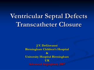
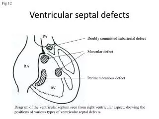
Fig 12. Ventricular septal defects. PA. Doubly committed subarterial defect. Muscular defect. RA. Perimembranous defect. RV. Diagram of the ventricular septum seen from right ventricular aspect, showing the positions of various types of ventricular septal defects. Fig 13.

Ventricular septal defects
E N D
Presentation Transcript
Fig 12 Ventricular septal defects PA Doubly committed subarterial defect Muscular defect RA Perimembranous defect RV Diagram of the ventricular septum seen from right ventricular aspect, showing the positions of various types of ventricular septal defects.
Fig 13 Small perimembranous VSD • Echocardiogram demonstrating shunting through a perimembranous ventricular septal defect. Parasternal long-axis view with the transducer tilted toward the right ventricular inflow. RV LV LA
Fig 14 Large perimembranous VSD with PH LV MPA RV AO LA RA Parasternal short axis view from same patient showing significantly dilated main pulmonary artery, indicating the presence of pulmonary hypertension Apical four chamber view demonstrating a large perimembranous inlet ventricular septal defect
Fig 15 Muscular VSD Apical five chamber view demonstrating a small muscular outlet ventricular septal defect (arrow) LV RV AO
Fig 16 Doubly committed VSD RV PA vsd Ao RA LA LA Modified Parasternal short axis view demonstrating the presence of doubly committed subarterial ventricular defect (arrow). Note there is no septal tissue between aortic and pulmonary valve.
Fig 17 Residual VSD Parasternal long-axis view showing a residual VSD. The echogenic patch on the septum suggests a previous repair. RV LV AO LA