Download
1 / 13
200 likes | 611 Views
Diagnostic Radiology: Congestive Heart Failure. Loren Morgan. Clinical Causes. HTN CAD Bad Valves (Aortic Stenosis, Mitral Stenosis) Cardiomyopathy L R Shunts. Classifications. Acute vs. Chronic Right Sided vs. Left Sided Low Output vs. High Output Systolic vs. Diastolic.

E N D
Diagnostic Radiology:Congestive Heart Failure Loren Morgan
Clinical Causes • HTN • CAD • Bad Valves (Aortic Stenosis, Mitral Stenosis) • Cardiomyopathy • LR Shunts
Classifications • Acute vs. Chronic • Right Sided vs. Left Sided • Low Output vs. High Output • Systolic vs. Diastolic
Clinical Presentation • Dyspnea • Orthopnea • Paroxysmal nocturnal dyspnea • Fatigue / Decreased exercise tolerance • Unexplained cough • Acute confusion, delirium • Abdominal symptoms (nausea, abdominal pain or distention) • Decreased food intake • Decline in functional status
Radiological Signs • Enlarged Heart Shadow (chronic CHF) • Cephalization • Kerley B Lines • Peri-bronchial Cuffing • Pulmonary Effusion • Pulmonary Interstitial Edema • Pulmonary Alveolar Edema
Cardiomegaly Cardiac width is larger than half trans-thoracic diameter. Cardiothoraccic rartio >0.5.
Cephalization Cephalization occurs due to increased flow to the apices of the lung as a result of increased pulmonary venous pressure. They stand out because there is more blood in them, the pressure is higher, and there may also be some edema surrounding them. For it to be correctly described the patient must be upright when the film is obtained.
Kerley B Lines These are short parallel lines at the lung periphery. These lines represent interlobular septa, which are usually less than 1 cm in length and parallel to one another at right angles to the pleural surface. They may be seen in any zone but are most frequently observed at the lung bases at the costophrenic angles on the PA radiograph, and in the substernal region on lateral radiographs
Peri-Bronchial Cuffing Occurs when excess fluid or mucus build up in the small airway passages of the lung causing localized patches of atelectasis. This causes the area around the bronchus to appear more prominent on an xray.
Pulmonary Effusions Fluid accumulation in the pleural space. Usually a trasudative effusion in CHF.
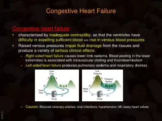
Pulm. Interstitial Edema Classical bat wing appearance is visible as bilateral hilar haze. Cardiogenic pulmonary edema occurs when the pulmonary capillary pressure exceeds 25 mm Hg. Above this level, interstitial pulmonary edema occurs which manifests clinicallly as shortness of breath and tachypnoea.
Pulm. Alveolar Edema As it progresses, alveolar edema occurs which manifests as dyspnoea with frothy blood stained sputum and bilateral basal crepitations. In more advanced stage, the crepitations (rales) extends through out the lung fields
References • www.meddean.lcu.edu • www.hcoa.org • www.cardiophile.org • www.learningradiology.com • www.emedicine.com/radiology