Download
1 / 182
1.82k likes | 1.83k Views
Learn about the lymphatic system, its protection against diseases, and specific defenses like lymphocytes. Explore lymph, lymphatic vessels, and immune system cells involved in immunity production.

E N D
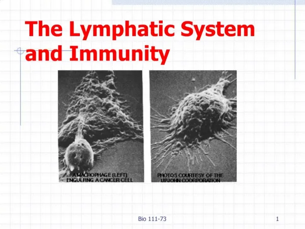
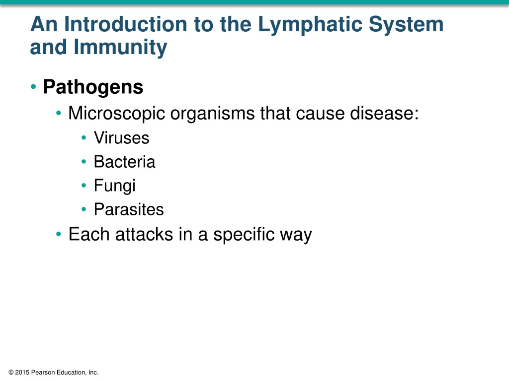
An Introduction to the Lymphatic System and Immunity • Pathogens • Microscopic organisms that cause disease: • Viruses • Bacteria • Fungi • Parasites • Each attacks in a specific way
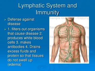
22-1 Overview of the Lymphatic System • The LymphaticSystem • Protects us against disease • Lymphatic system cells respond to: • Environmental pathogens • Toxins • Abnormal body cells, such as cancers
22-1 Overview of the Lymphatic System • Specific Defenses • Lymphocytes • Part of the immuneresponse • Identify, attack, and develop immunity • To a specific pathogen
22-1 Overview of the Lymphatic System • The ImmuneSystem • Immunity • The ability to resist infection and disease • All body cells and tissues involved in production of immunity • Not just lymphatic system
22-1 Overview of the Lymphatic System • Nonspecific Defenses • Block or attack any potential infectious organism • Cannot distinguish one attack from another
22-2 Structures of Body Defenses • Organization of the Lymphatic System • Lymph • A fluid similar to plasma but does not have plasma proteins • Lymphaticvessels (lymphatics) • Carry lymph from peripheral tissues to the venous system • Lymphoidtissues and lymphoidorgans • Lymphocytes, phagocytes, and other immune system cells
Lymph Lymphocyte Figure 22-1 An Overview of the Lymphatic System (Part 1 of 2). Lymphatic Vesselsand Lymph Nodes Cervical lymph nodes Thoracic duct Right lymphatic duct Lymphoid Tissuesand Organs Axillary lymph nodes Lymphatics ofmammary gland Tonsil Thymus Cisterna chyli Spleen Lymphatics of upper limb Mucosa-associatedlymphoid tissue(MALT) in digestive,respiratory, urinary,and reproductivetracts Lumbar lymph nodes
Lymphatic Vesselsand Lymph Nodes Lymphoid Tissuesand Organs Pelvic lymph nodes Appendix Red bone marrow Figure 22-1 An Overview of the Lymphatic System (Part 2 of 2). Inguinal lymph nodes Lymphatics of lower limb
Arteriole Lymphaticcapillary Smoothmuscle Endothelialcells Figure 22-2a Lymphatic Capillaries. Venule Interstitialfluid Lymphflow Blood capillaries Areolar tissue a The interwoven network formed by blood capillariesand lymphatic capillaries. Arrows indicate themovement of fluid out of blood capillaries and the net flow of interstitial fluid and lymph.
Lymphocyte Incompletebasementmembrane Lymphflow To largerlymphatics Figure 22-2b Lymphatic Capillaries. Areolartissue Interstitial fluid Plasma Interstitialfluid Lymphaticcapillary Bloodcapillary b A sectional view indicating the movement of fluidfrom the plasma, through the tissues as interstitialfluid, and into the lymphatic system as lymph.
Artery Vein Figure 22-3a Lymphatic Vessels and Valves. Lymphaticvessel Vein Artery Lymphaticvessel a Towardvenoussystem Lymphaticvalve From lymphaticcapillaries A diagrammatic view of areolar connective tissue containing small blood vessels and a lymphatic vessel. The cross-sectional view emphasizes theirstructural differences.
Lymphaticvalve Figure 22-3b Lymphatic Vessels and Valves. Lymphaticvessel Lymphatic vessel and valve LM × 63 Like valves in veins, each lymphatic valve consists of a pair of flaps that permit movement of fluid in only one direction. b
Left internal jugular vein Brachiocephalicveins Left jugular trunk Right internal jugular vein Right jugular trunk Thoracic duct Left subclavian trunk Right lymphatic duct Right subclavian trunk Left bronchomediastinaltrunk Right subclavian vein Left subclavianvein Right bronchomediastinaltrunk Superior vena cava (cut) First rib (cut) Figure 22-4 The Relationship between the Lymphatic Ducts and the Venous System. Azygos vein Highestintercostalvein Rib (cut) Thoracicduct Drainage of rightlymphaticduct Drainageof thoracicduct Thoraciclymph nodes Hemiazygosvein Parietalpleura (cut) Diaphragm Cisterna chyli Inferior vena cava (cut) Intestinal trunk Right lumbar trunk Left lumbar trunk a The thoracic duct carrieslymph originating in tissuesinferior to the diaphragmand from the left side of theupper body. The smaller rightlymphatic duct carries lymphfrom the rest of the body. The thoracic duct empties into the left subclavianvein. The right lymphatic duct empties into theright subclavian vein. b
Drainage of rightlymphaticduct Drainageof thoracicduct Figure 22-4a The Relationship between the Lymphatic Ducts and the Venous System. a The thoracic duct carrieslymph originating in tissuesinferior to the diaphragmand from the left side of theupper body. The smaller rightlymphatic duct carries lymphfrom the rest of the body.
22-2 Structures of Body Defenses • Lymphedema • Blockage of lymph drainage from a limb • Causes severe swelling • Interferes with immune system function • Lymphocytes • Make up 20–40 percent of circulating leukocytes • Most are stored, not circulating
22-2 Structures of Body Defenses • Types of Lymphocytes • T cells • Thymus-dependent • B cells • Bone marrow-derived • NK cells • Natural killer cells
22-2 Structures of Body Defenses • T Cells • Make up 80 percent of circulating lymphocytes • Main Types of T Cells • Cytotoxic T (TC) cells • Memory T cells • Helper T (TH) cells • Suppressor T (TS) cells
22-2 Structures of Body Defenses • Cytotoxic T Cells • Attack cells infected by viruses • Produce cell-mediated immunity • Memory T Cells • Formed in response to foreign substance • Remain in body to give “immunity” • Helper T Cells • Stimulate function of T cells and B cells
22-2 Structures of Body Defenses • Suppressor T Cells • Inhibit function of T cells and B cells • Regulatory T Cells • Are helper and suppressor T cells • Control sensitivity of immune response
22-2 Structures of Body Defenses • Other T Cells • Inflammatory T cells • Suppressor/inducer T cells • B Cells • Make up 10–15 percent of circulating lymphocytes • Differentiate (change) into plasmacells • Plasma cells • Produce and secrete antibodies (immunoglobulin proteins)
22-2 Structures of Body Defenses • Antigens • Targets that identify any pathogen or foreign compound • Immunoglobulins (Antibodies) • The binding of a specific antibody to its specific target antigen initiates antibody-mediatedimmunity
22-2 Structures of Body Defenses • Antibody-Mediated Immunity • A chain of events that destroys the target compound or organism • NaturalKiller (NK) Cells • Also called largegranularlymphocytes • Make up 5–10 percent of circulating lymphocytes • Responsible for immunologicalsurveillance • Attack foreign cells, virus-infected cells, and cancer cells
CAR T-cell therapy; drawing of blood being removed from a vein in a patient’s arm to get T cells. Also shown is a special receptor called a chimeric antigen receptor (CAR) being made in the laboratory; the gene for CAR is inserted into the T cells and then millions of CAR T cells are grown. Drawing also shows the CAR T cells being given to the patient by infusion and binding to antigens on the cancer cells and killing them.CAR T-cell therapy. A type of treatment in which a patient’s T cells (a type of immune cell) are changed in the laboratory so they will bind to cancer cells and kill them. Blood from a vein in the patient’s arm flows through a tube to an apheresis machine (not shown), which removes the white blood cells, including the T cells, and sends the rest of the blood back to the patient. Then, the gene for a special receptor called a chimeric antigen receptor (CAR) is inserted into the T cells in the laboratory. Millions of the CAR T cells are grown in the laboratory and then given to the patient by infusion. The CAR T cells are able to bind to an antigen on the cancer cells and kill them.
Classes of Lymphocytes subdivided into T Cells Approximately 80% of circulatinglymphocytes are classified as T cells. Figure 22-5 Classes of Lymphocytes (Part 1 of 2). differentiate into HelperT Cells CytotoxicT Cells SuppressorT Cells Memory T Cells Cytotoxic T cellsattack foreign cellsor body cellsinfected by viruses. Helper T cells stimulate theactivation andfunction ofboth T cellsand B cells. Suppressor Tcells inhibitthe activationand functionof both T cells and B cells. Memory T cellsare a subsetof T cells that respond to apreviouslyencounteredantigen.
Classes of Lymphocytes subdivided into B Cells NK Cells NK cellsmake up theremaining5–10% ofcirculatinglymphocytes. B cells make up10–15% ofcirculatinglymphocytes. Figure 22-5 Classes of Lymphocytes (Part 2 of 2). Plasma Cells When stimulated, Bcells can differentiateinto plasma cells,which produce andsecrete antibodies.
22-2 Structures of Body Defenses • Lymphocyte Production • Also called lymphopoiesis, involves: • Bone marrow • Thymus • Peripheral lymphoid tissues • Hemocytoblasts • In bone marrow, divide into two types of lymphoid stem cells
22-2 Structures of Body Defenses • Lymphoid Stem Cells • Group 1 • Remains in bone marrow and develop with help of stromalcells • Produces B cells and natural killer cells • Group 2 • Migrates to thymus • Produces T cells in environment isolated by blood–thymusbarrier
Red Bone Marrow One group of stem cells remains inthe red bone marrow, producingdaughter cells that mature into NKcells and B cells. Multipotenthemopoieticstem cell Figure 22-6 The Origin and Distribution of Lymphocytes (Part 1 of 3). Interleukin-7 Lymphoid stem cells Lymphoid stem cells B cells NK cells
Thymus The second group of stemcells migrates to the thymus,where subsequent divisionsproduce daughter cells thatmature into T cells. Figure 22-6 The Origin and Distribution of Lymphocytes (Part 2 of 3). Thymichormones Lymphoid stem cells Production,selection, anddifferentiatiionof T cells Mature T cells Mature T cells
Peripheral Tissues All three typesof lymphocytes circulatethroughout thebody in thebloodstream,establishingimmunity. Antibody-mediated immunity Immune surveillance Cell-mediated immunity Figure 22-6 The Origin and Distribution of Lymphocytes (Part 3 of 3). One type of mature Tcell, called cytotoxicT cells, plays arole in cell-mediatedimmunity. Thesecells attack anddestroy foreigncells or bodycells infectedby viruses. When stimulated, Bcells can differentiate into plasma cells,which produce andsecrete antibodies.These antibodiesattach to pathogens.This starts achain reactionthat leads tothe destructionof the pathogen. NK cells attackforeign cells, bodycells infected byviruses, andcancer cells.They secretechemicals thatlyse the plasmamembrane ofthe abnormalcells. B cell CytotoxicT cell NK cells Abnormalcell Abnormalcell Plasma cell Antibodies Cell destroyed Cell destroyed
22-2 Structures of Body Defenses • T Cells and B Cells • Migrate throughout the body • To defend peripheral tissues • Retaining their ability to divide • Is essential to immune system function
22-2 Structures of Body Defenses • Differentiation • B cells differentiate • With exposure to hormone called cytokine (interleukin-7) • T cells differentiate • With exposure to several thymic hormones
22-2 Structures of Body Defenses • LymphoidTissues • Connective tissues dominated by lymphocytes • LymphoidNodules • Areolar tissue with densely packed lymphocytes • Germinal center contains dividing lymphocytes • Lab
22-2 Structures of Body Defenses • The Lymphatic System and Body Defenses • Body defenses provide resistance to fight infection, illness, and disease • Two categories of defenses • Innate (nonspecific) immunity • Adaptive (specific) immunity
22-2 Structures of Body Defenses • Innate (Nonspecific) Immunity • Always works the same way • Against any type of invading agent • Nonspecificresistance • Adaptive (Specific) Immunity • Protects against specific pathogens • Depends on activities of lymphocytes • Specificresistance (immunity) • Develops after exposure to environmental hazards
22-3 Nonspecific Defenses • Seven Major Categories of Innate (Nonspecific) Immunity • Physicalbarriers • Phagocytes • Immunesurveillance • Interferons • Complement • Inflammatoryresponse • Fever
22-3 Nonspecific Defenses • Physical Barriers • Keep hazardous materials outside the body • Phagocytes • Attack and remove dangerous microorganisms • Immune Surveillance • Constantly monitors normal tissues • With natural killer cells (NK cells)
22-3 Nonspecific Defenses • Interferons • Chemical messengers that trigger production of antiviral proteins in normal cells • Antiviral proteins • Do not kill viruses • Block replication in cell • Complement • System of circulating proteins • Assists antibodies in destruction of pathogens
22-3 Nonspecific Defenses • Inflammatory Response • Localized, tissue-level response that tends to limit spread of injury or infection • Fever • A high body temperature • Increases body metabolism • Accelerates defenses • Inhibits some viruses and bacteria
Innate Defenses Physical barriers Duct of eccrinesweat gland Secretions Hair keep hazardousorganisms andmaterials outsidethe body. Epithelium Figure 22-11 Innate Defenses (Part 1 of 2). Phagocytes engulf pathogensand cell debris. Fixedmacrophage Freemacrophage Monocyte Eosinophil Neutrophil Immune surveillance is the destruction ofabnormal cells by NKcells in peripheral tissues. Lysedabnormalcell Naturalkiller cell Interferons are chemical messengersthat coordinate thedefenses against viralinfections. Interferons released by activatedlymphocytes, macrophages, orvirus-infected cells
Innate Defenses Complement is a system of circulatingproteins that assistantibodies in thedestruction of pathogens. Lysedpathogen Figure 22-11 Innate Defenses (Part 2 of 2). Complement Inflammation Blood flow increased Phagocytes activated Capillary permeability increased Complement activated Clotting reaction walls off region Regional temperature increased Adaptive defenses activated is a localized, tissue-levelresponse that tends tolimit the spread of aninjury or infection. Mast cell Fever is an elevation of bodytemperature that acceleratestissue metabolism andbody defenses. Body temperature rises above 37.2C inresponse to pyrogens
22-3 Nonspecific Defenses • Physical Barriers • Outer layer of skin • Hair • Epithelial layers of internal passageways • Secretions that flush away materials • Sweat glands, mucus, and urine • Secretions that kill or inhibit microorganisms • Enzymes, antibodies, and stomach acid
22-3 Nonspecific Defenses • Two Classes of Phagocytes • Microphages • Neutrophils and eosinophils • Leave the bloodstream • Enter peripheral tissues to fight infections
22-3 Nonspecific Defenses • Two Classes of Phagocytes • Macrophages • Large phagocytic cells derived from monocytes • Distributed throughout body • Make up monocyte–macrophage system (reticuloendothelialsystem)
22-3 Nonspecific Defenses • Activated Macrophages • Respond to pathogens in several ways • Engulf pathogen and destroy it with lysosomal enzymes • Bind to pathogen so other cells can destroy it • Destroy pathogen by releasing toxic chemicals into interstitial fluid
22-3 Nonspecific Defenses • Two Types of Macrophages • Fixedmacrophages • Also called histiocytes • Stay in specific tissues or organs • For example, dermis and bone marrow • Freemacrophages • Also called wanderingmacrophages • Travel throughout body
22-3 Nonspecific Defenses • Special Histiocytes • Microglia found in central nervous system • Kupffercells found in liver sinusoids • Free Macrophages • Special free macrophages • Alveolarmacrophages (phagocyticdustcells)
22-3 Nonspecific Defenses • Movement and Phagocytosis • All macrophages: • Move through capillary walls (emigration) • Are attracted or repelled by chemicals in surrounding fluids (chemotaxis) • Phagocytosis begins: • When phagocyte attaches to target (adhesion) • And surrounds it with a vesicle