Download
1 / 42
450 likes | 965 Views
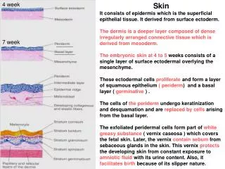
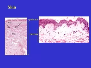
Staphylococcus aureus is the major pathogen for the skin of mice and rats. Staphyloccus aureus is a gram-positive coccus that typically grows in clusters . It commonly inhabits the skin, skin glands, nasopharynx, and intestine of many host species. Skin.

E N D
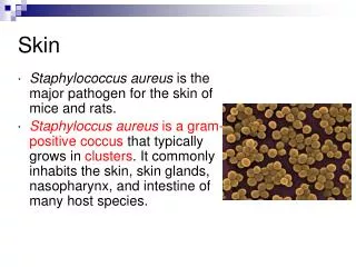
Staphylococcus aureus is the major pathogen for the skin of mice and rats. Staphyloccus aureus is a gram-positive coccus that typically grows in clusters. It commonly inhabits the skin, skin glands, nasopharynx, and intestine of many host species. Skin
Staph. aureus is a common commensal of many species. It also is common in the environment. Human carriers can be an important source of infection for rodent colonies
Staph. aureus is associated mostly with dermatitis in the form of pyoderma or ulcerative dermatitis. The face, shoulders, neck, and ears are most commonly affected.
Abscesses and granulomas, most commonly affecting the face and tissues around the base of the tail, also occur and can be associated with fight wounds Staphylococcus aureus
The best methods of control are improved sanitation, frequent sanitizing of cages and other equipment, and elimination of equipment that could cause skin injury Topical treatment with Nolvasan BID Staphylococcus aureus
Sialodacryoadenitis virus (SDAV), another coronavirus (Parainfluenza 1), is one of the most common viruses found in laboratory rats and mice. It is highly contagious, and is spread by direct contact with infected animals or by respiratory aerosol SDAV (Sendai Virus) SV
The incubation period for SDAV less than 1 week. In naive populations, a sudden high incidence of overt disease with sneezing, porphyrin-stained nasal and ocular discharges (as seen in this image), cervical edema, corneal ulceration, and keratoconus may be the first indications of a problem SV
This image shows swollen submandibular salivary glands (arrows) in a mouse with SV. SV has tissue tropism for the submaxillary and parotid salivary, exorbital, Harderian, and intraorbital lacrimal glands Sendai Virus
In rats and mice, few gross morphologic lesions are seen in uncomplicated Sendai infections. The lungs can be focally reddened and atelectatic with serous fluid visible in the pleural and pericardial cavities.
Mouse hepatitis virus (MHV) and sialodacryoadenitis virus (SDAV) are frequently encountered coronaviruses of mice and rats Some primarily infect the gastrointestinal system; some the respiratory tract; some the brain Mouse Hepatitis Virus (MHV)
Mice (Mus musculus) are the only natural hosts. MHV is extremely contagious with prevalence rates exceeding 80% in outbreaks. Active infection lasts 2-3 weeks, during which mice shed the virus in gastrointestinal and respiratory excretions. Direct contact with shedding mice, contaminated cell lines, fomites, or airborne particles are the important routes of viral transmission. MHV
can produce nonspecific clinical signs in naïve*, juvenile mice, such as runting, as shown here, or failure to thrive MHV not previously subjected to experimentation or a particular experimental situation <made the test with naive mouse>
This image shows large coalescing cream-colored friable foci (arrow) of necrosis that result when acute multifocal hepatitis progresses to chronic active hepatitis in nude mice. Gross pathology in immune-incompetent mice is more generalized and progressive than in immune-competent mice. MHV
Enterotropic versus respiratory strains Extremely contagious by many routes Nursing pups: diarrhea and mortality Weanlings: Obstipation Adults: Hunched posture, weight loss, rough hair coat, variable mortality No latent infections- stop breeding ELISA Mouse Hepatitis Virus
Diagnosis of latent infections is dependent on the histologic demonstration of large, multinucleate syncytial cells (arrow) in the liver, brain, or mucosal epithelium of the intestine Mouse Hepatitis Virus
Rotavirus, another genus of the family Reoviridae, is associated with clinical disease. Rotaviruses affecting mice and rats respectively are mouse rotavirus, a group-A rotavirus associated with the syndrome, epizootic diarrhea of infant mice (EDIM) Rotavirus (EDIM)
Epizootic Diarrhea of Infant Mice (EDIM) Most susceptible: birth to 17 days of age Fecal-oral transmission Yellow, watery diarrhea in 14-17 day old pups Death with a full stomach (versus reovirus), shortened intestinal villi Unapparent viral carriers ELISA Rotavirus
Neonatal diarrhea is the most prominent sign. Watery yellow stool accumulates around the anus and tailbase, soiling the coats of neonatal pups and their dams. Pups appear stunted and lethargic and have distended abdomens. Mortality rates are low EDIM
Common Transmitted by direct contact May be subclinical, or develop pruritus, scruffy coat, patchy alopecia, self-trauma, pyoderma Typically found on back and head Mist with ivermectin (0.1%) for 3 weekly treatments Myobia musculi, Radfordia affinis, and Myocoptes musculinus
Myobia musculi lesions in a pet white mouse, characteristic of hypersensitivity to the mites. Intense pruritus, often directed at the neck and ears, leads to self-mutilation. Early lesions consist of subtle hair thinning on the dorsal neck and shoulders Acariasis??? Myobia musculi
Myobia musculi Single claw
Radfordia affinis Two claws
Common Fecal-oral transmission Usually subclinical, but may cause rectal prolapse Fecal floatation (Aspiculuris tetraptera) or tape test (Syphacia obvelata) Ivermectin misting (others more labor intensive) Clean environment well Pinworms
Common tapeworms among pet mice Roaches, beetles, & fleas = intermediate hosts R. nana: also transmitted directly, or by autoinfection (retroinfection) Usually subclinical infection Rodentolepis nana and Hymenolepis diminuta
Often find proglottids in feces instead of individual eggs Zoonotic (more commonly R. nana because of direct transmission) Praziquantel is effective Rodentolepis nana and Hymenolepis diminuta
Rodentolepis nana Polar filaments
Most common tumor of mice Mice have 3 pair of thoracic and 2 pair of abdominal mammary glands- glandular tissue may be found up around the body to the dorsum Poor prognosis- anaplastic and very invasive Mammary Adenocarcinoma
Animal model of trichotillomania MOBS (“Move Over Buddy” Syndrome) Alopecia (R.O. ectoparasites, dermatophytes, endocrinopathy) Well demarcated area of alopecia without dermatitis- exposed skin appears normal Commonly involves hair over the nasal and orbital regions, or over the dorsal cervical area Separate out barber Barbering
Males fight and abuse females Bites often found on face, back, and genital area May abscess Nolvasan (+ lance abscesses) Separate offenders Provide enrichment Bite Wounds
Genetic predisposition (autosomal recessive) Incisors hypsodont Inanition, starvation Trim teeth with nail clippers (no “scissor” action) Do not breed these mice Malocclusion