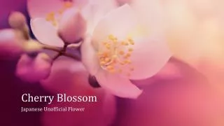
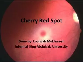
Cherry Red Spot
Cherry Red Spot. Done by: Loulwah Mukharesh Intern at King Abdulaziz University. Etiology. The characteristic pale hue heavy deposition of lipid, sphingolipid , or oligosaccharides in the ganglionic cells of the retina at the macula .

Cherry Red Spot
E N D
Presentation Transcript
Cherry Red Spot Done by: Loulwah Mukharesh Intern at King Abdulaziz University
Etiology • The characteristic pale hueheavydeposition of lipid, sphingolipid, or oligosaccharides in the ganglionic cells of the retina at the macula. • In the center of the pale region lies the foveal pit which lacks ganglion cellscontinuesto retain its reddish appearance.
History • 1887 by Bernard Sachs “arrested development with special reference to its cortical pathology”. • Neuropathologic examination confirmed lipid storage disease in the brain in a child. • This child was also seen by Herman Knapp, an ophthalmologist who practiced in NY and Berlin and described the retinal features of this child at an ophthalmology meeting at Heidelberg and was the first to use the term “cherry-red color” to describe the fovea. • Subsequently the child was found to have Tay Sachs disease. • Knapp had initially thought that the cherry-red spot was a benign finding but later realized its grave implications.
Differential Diagnosis • It is also seen in other neurometabolicdiseases as well as in central retinal artery occlusion.
Controversy • The cherry-red spot may become less prominent over time, concurrent with loss of the affected peri-macular ganglion cells. • Because the cherry-red appearance of the retina is characteristic only of Caucasians, and because the retinal complexion differs based on ethnicity, it was suggested that the term “peri-fovealwhite patch” may be more appropriate than cherry-red spot.
References • “Cherry-red spot” or “perifoveal white patch”? Luis H. Ospina et al., Can J Ophthalmol 2005;40:609–10 • The “Cherry Red” Spot, Jacqueline A. Leavitt et al. Pediatric Neurology Volume 37 (1) Elsevier – Jul 1, 2007 • UpToDate