Download
1 / 1
10 likes | 117 Views
Solid State NMR Investigation of Toxic Particles Formed by the Alzheimer’s Amyloid- b Protein. Robert Wallace, Wakulla High School, Crawfordville, FL 32327 Joel Falk, Acquia Madre Elementary School, Sante Fe, NM 87505

E N D
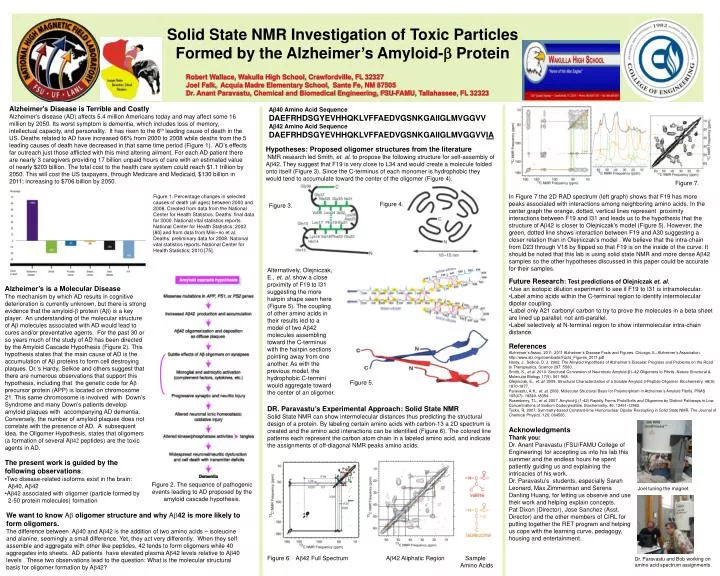
Solid State NMR Investigation of Toxic Particles Formed by the Alzheimer’s Amyloid-b Protein Robert Wallace, Wakulla High School, Crawfordville, FL 32327 Joel Falk, Acquia Madre Elementary School, Sante Fe, NM 87505 Dr. Anant Paravastu, Chemical and Biomedical Engineering, FSU-FAMU, Tallahassee, FL 32323 Alzheimer's Disease is Terrible and Costly Alzheimer's disease (AD) affects 5.4 million Americans today and may affect some 16 million by 2050. Its worst symptom is dementia, which includes loss of memory, intellectual capacity, and personality. It has risen to the 6th leading cause of death in the US. Deaths related to AD have increased 66% from 2000 to 2008 while deaths from the 5 leading causes of death have decreased in that same time period (Figure 1). AD’s effects far outreach just those afflicted with this mind altering ailment. For each AD patient there are nearly 3 caregivers providing 17 billion unpaid hours of care with an estimated value of nearly $203 billion. The total cost to the health care system could reach $1.1 trillion by 2050. This will cost the US taxpayers, through Medicare and Medicaid, $130 billion in 2011; increasing to $706 billion by 2050. Ab40 Amino Acid Sequence DAEFRHDSGYEVHHQKLVFFAEDVGSNKGAIIGLMVGGVV Ab42 Amino Acid Sequence DAEFRHDSGYEVHHQKLVFFAEDVGSNKGAIIGLMVGGVVIA Hypotheses: Proposed oligomer structures from the literature NMR research led Smith, et. al. to propose the following structure for self-assembly of Ab42. They suggest that F19 is very close to L34 and would create a molecule folded onto itself (Figure 3). Since the C-terminus of each monomer is hydrophobic they would tend to accumulate toward the center of the oligomer (Figure 4). Figure 7. In Figure 7 the 2D RAD spectrum (left graph) shows that F19 has more peaks associated with interactions among neighboring amino acids. In the center graph the orange, dotted, vertical lines represent proximity interactions between F19 and I31 and leads us to the hypothesis that the structure of Ab42 is closer to Olejniczak’s model (Figure 5). However, the green, dotted line shows interaction between F19 and A30 suggesting a closer relation than in Olejniczak’s model . We believe that the intra-chain from D23 through V18 by flipped so that F19 is on the inside of the curve. It should be noted that this lab is using solid state NMR and more dense Ab42 samples so the other hypotheses discussed in this paper could be accurate for their samples. Figure 1. Percentage changes in selected causes of death (all ages) between 2000 and 2008. Created from data from the National Center for Health Statistics. Deaths: final data for 2000. National vital statistics reports. National Center for Health Statistics; 2002 [80] and from data from Mini~no et al. Deaths: preliminary data for 2008. National vital statistics reports. National Center for Health Statistics; 2010 [75]. Figure 4. Figure 3. Alternatively, Olejniczak, E., et. aI. show a close proximity of F19 to I31 suggesting the more hairpin shape seen here (Figure 5). The coupling of other amino acids in their results led to a model of two Ab42 molecules assembling toward the C-terminus with the hairpin sections pointing away from one another. As with the previous model, the hydrophobic C-termini would aggregate toward the center of an oligomer. • Future Research: Test predictions of Olejniczak et. al. • Use an isotopic dilution experiment to see if F19 to I31 is intramolecular. • Label amino acids within the C-terminal region to identify intermolecular dipolar coupling. • Label only A21 carbonyl carbon to try to prove the molecules in a beta sheet are lined up parallel, not anti-parallel. • Label selectively at N-terminal region to show intermolecular intra-chain distance. • Alzheimer’s is a Molecular Disease • The mechanism by which AD results in cognitive deterioration is currently unknown, but there is strong evidence that the amyloid-b protein (Ab) is a key player. An understanding of the molecular structure of Abmolecules associated with AD would lead to cures and/or preventative agents. For the past 30 or so years much of the study of AD has been directed by the Amyloid Cascade Hypothesis (Figure 2). This hypothesis states that the main cause of AD is the accumulation of Ab proteins to form cell destroying plaques. Dr.’s Hardy, Selkoe and others suggest that there are numerous observations that support this hypothesis, including that the genetic code for Ab precursor protein (APP) is located on chromosome 21. This same chromosome is involved with Down’s Syndrome and many Down’s patients develop amyloid plaques with accompanying AD dementia. Conversely, the number of amyloid plaques does not correlate with the presence of AD. A subsequent idea, the Oligomer Hypothesis, states that oligomers (a formation of several Ab42 peptides) are the toxic agents in AD. • The present work is guided by the following observations: • Two disease-related isoforms exist in the brain: Ab40, Ab42 • Ab42 associated with oligomer (particle formed by • 2-50 protein molecules) formation References Alzheimer’s Assoc. 2011. 2011 Alzheimer’s Disease Facts and Figures. Chicago, IL: Alzheimer’s Association. http://www.alz.org/downloads/Facts_Figures_2011.pdf Hardy, J., Selkoe, D. J. 2002. The Amyloid Hypothesis of Alzheimer’s Disease: Progress and Problems on the Road to Therapeutics, Science 297: 5580. Smith, S., et al. 2010. Structural Conversion of Neurotoxic Amyloid-β1–42 Oligomers to Fibrils. Nature Structural & Molecular Biology 17(5): 561-568. Olejniczak, E., et. aI. 2009. Structural Characterization of a Soluble Amyloid b-Peptide Oligomer. Biochemistry. 48(9): 1870-1877. Paravastu, A.K., et. aI. 2008. Molecular Structural Basis for Polymorphism in Alzheimer’s Amyloid Fibrils. PNAS 105(47): 18349-18354 Rosenberry, T.L, et al. 2007. Amyloid-b (1-42) Rapidly Forms Protofibrils and Oligomers by Distinct Pathways in Low Concentrations of Sodium Dodecylsulfate. Biochemistry. 46: 12451-12462. Tycko, R. 2007. Symmetry-based Constant-time Homonuclear Dipolar Recoupling in Solid State NMR. The Journal of Chemical Physics. 126: 064506. Figure 5. DR. Paravastu’s Experimental Approach: Solid State NMR Solid State NMR can show intermolecular distances thus predicting the structural design of a protein. By labeling certain amino acids with carbon-13 a 2D spectrum is created and the amino acid interactions can be identified (Figure 6). The colored line patterns each represent the carbon atom chain in a labeled amino acid, and indicate the assignments of off-diagonal NMR peaks amino acids. Acknowledgments Thank you: Dr. Anant Paravastu (FSU/FAMU College of Engineering) for accepting us into his lab this summer and the endless hours he spent patiently guiding us and explaining the intricacies of his work. Dr. Paravastu’s students, especially Sarah Leonard, Max Zimmerman and Serena Danting Huang, for letting us observe and use their work and helping explain concepts. Pat Dixon (Director), Jose Sanchez (Asst. Director) and the other members of CIRL for putting together the RET program and helping us cope with the learning curve, pedagogy, housing and entertainment. Figure 2. The sequence of pathogenic events leading to AD proposed by the amyloid cascade hypothesis. Joel tuning the magnet. We want to know Ab oligomer structure and why Ab42 is more likely to form oligomers. The difference between Ab40 and Ab42 is the addition of two amino acids – isoleucine and alanine, seemingly a small difference. Yet, they act very differently. When they self assemble and aggregate with other like peptides, 42 tends to form oligomers while 40 aggregates into sheets. AD patients have elevated plasma Ab42 levels relative to Ab40 levels . These two observations lead to the question: What is the molecular structural basis for oligomer formation by Ab42? Figure 6. Ab42 Full Spectrum Ab42 Aliphatic Region Sample Amino Acids Dr. Paravastu and Bob working on amino acid spectrum assignments.