Inflammation
Inflammation. Zhu keqing 竺可青 Pathology Department Zhejiang University School of Medicine zhukeqing@zju.edu.cn 2012-10. 炎症基本概念 炎症原因 炎症基本病理变化 炎症介质 炎症结局 炎症常见组织类型. Basic change. Alteration 变质 Exudation 渗出 Proliferation 增生. 炎症的组织学类型 Morphologic Patterns of Acute Inflammation.

Inflammation
E N D
Presentation Transcript
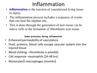
Inflammation Zhu keqing 竺可青 Pathology Department Zhejiang University School of Medicine zhukeqing@zju.edu.cn 2012-10
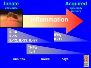
炎症基本概念 • 炎症原因 • 炎症基本病理变化 • 炎症介质 • 炎症结局 • 炎症常见组织类型
Basic change • Alteration 变质 • Exudation 渗出 • Proliferation 增生
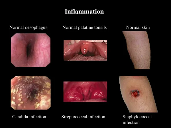
炎症的组织学类型Morphologic Patterns of Acute Inflammation • 以组织细胞变性、坏死为主的炎症称为变质性炎。 • 急性重症肝炎、流行性乙型脑炎、阿米巴原虫肝、白喉中毒性心肌炎 • 以浆液、纤维蛋白原和中性粒细胞渗出为主的炎症称渗出性炎 • 浆液性炎、纤维素性炎、化脓性炎、出血性炎 • SEROUS INFLAMMATION • FIBRINOUS INFLAMMATION • SUPPURATIVE OR PURULENT INFLAMMATION • ULCERS
浆液性炎SEROUS INFLAMMATION • 浆液渗出为主 • 渗出的蛋白以小分子白蛋白为主 • 伴少量纤维蛋白和炎症细胞 • 血管壁损伤轻 • 少量渗出可完全吸收,不留痕迹 • 大量渗出引起压迫和水肿 • 发生与皮肤、粘膜、浆液和疏松结缔组织
Serous inflammation. Low-power view of a cross-section of a skin blister showing the epidermis separated from the dermis by a focal collection of serous effusion.
纤维蛋白性炎FIBRINOUS INFLAMMATION • 纤维蛋白渗出为主 • 血管壁损伤较重 • 发生于粘膜、浆膜和肺组织 1. 假膜性炎(白喉、菌痢) • 发生在粘膜面的纤维蛋白性炎 • 假膜:纤维蛋白性、中性粒细胞、坏死粘膜上皮、病原微生物 2. 绒毛心 • 发生在心外膜的纤维蛋白性炎
Fibrinous pericarditis. Deposits of fibrin on the pericardium.
Fibrinous pericarditisA pink meshwork of fibrin exudate (F) overlies the pericardial surface
Seen here is vasodilation with exudation that has led to an outpouring of fluid with fibrin into the alveolar spaces, along with PMN's.
Here is an example of the fibrin mesh in fluid with PMN's that has formed in the area of acute inflammation. It is this fluid collection that produces the "tumor" or swelling aspect of acute inflammation.
This yellow-green exudate on the surface of an inflamed, hyperemic (erythematous) bowel mucosa consists of many neutrophils along with fibrin and amorphous debris from dying cells. 细菌性痢疾
Membrane of diphtheria lying within a transverse bronchus (A) and forming a perfect cast (removed from the lung) of the branching respiratory tree (B).
Here, the pericardial cavity has been opened to reveal a fibrinous pericarditis with strands of stringy pale fibrin between visceral and parietal pericardium.
Microscopically, the fibrinous exudate is seen to consist of pink strands of fibrin jutting from the pericardial surface at the upper left.
化脓性炎SUPPURATIVE OR PURULENT INFLAMMATION • 化脓性炎:大量中性粒细胞渗出为主伴组织坏死和脓液形成。 • 化脓suppuration:中性粒细胞和坏死组织崩解,释放蛋白酶,使坏死组织溶解液化成液状物的过程。 • 所形成的液状物称脓液pus。 • 变性坏死的白细胞称浓细胞。
1. 脓肿 abscess---局限性化脓伴浓腔形成。 • 病灶局限 • 主要由金黄色葡萄球菌引起 • 浓腔内有脓液 • 早期壁不规则,后期壁厚为大量肉芽组织 • 疖和痈为典型皮肤脓肿 2. 蜂窝织炎 phlegmonous inflammation • 弥漫性化脓性炎 • 主要由溶血性链球菌引起 • 链球菌分泌透明质酸酶和链激酶 • 大量中性粒细胞弥漫浸润 • 与正常组织分界不清 • 坏死不明显 • 常见于疏松组织:皮下、肌肉、阑尾 3. 表面化脓和积浓 • 表面化脓---粘膜组织的化脓性炎仅累及粘膜层。 • 积浓---浓性渗出物不能排出,聚集在输卵管、胆囊或胸腹腔内。
Here is a purulent exudate in which the exuded fluid also contains a large number of acute inflammatory cells. Thus, the yellowish fluid in this opened pericardial cavity is a purulent exudate.
Suppurative inflammationA, A subcutaneous bacterial abscess with collections of pus. B, The abscess contains neutrophils, edema fluid, and cellular debris
A purulent exudate is seen beneath the meninges in the brain of this patient with acute meningitis from Streptococcus pneumoniae infection. The exudate obscures the sulci.
The abdominal cavity is opened at autopsy here to reveal an extensive purulent peritonitis that resulted from rupture of the colon. A thick yellow exudate coats the peritoneal surfaces.
溃疡 ulcer • 皮肤粘膜的浅表脓肿,向表面破溃形成缺损。 • 窦道 sinus • 深部脓肿向体表或体腔穿破,一端为盲端的排浓管道。 • 瘘管 fistula • 有两个或两个以上的排浓管道。 • 空洞 cavity • 内脏器官脓肿的脓液经自然管道排出,形成空腔。
The morphology of an ulcer. A, A chronic duodenal ulcer. B, Low-power cross-section of a duodenal ulcer crater with an acute inflammatory exudate in the base.
出血性炎 Hemorrhagic inflamation • 血管损伤严重,渗出物中大量红细胞 • 流行性出血热、钩端螺旋体病、鼠疫
Chronic inflammation • Chronic inflammation is considered to be inflammation of prolonged duration (weeks or months) in which active inflammation, tissue destruction, and attempts at repair are proceeding simultaneously. • Although it may follow acute inflammation, as described earlier, chronic inflammation frequently begins insidiously, as a low-grade, smoldering, often asymptomatic response. • This latter type of chronic inflammation is the cause of tissue damage in some of the most common and disabling human diseases, such as rheumatoid arthritis, atherosclerosis, tuberculosis, and chronic lung diseases. • 以增生变化为主的炎症称为增生性炎,多为慢性炎。 • 包括非特异性增生性炎和特异性增生性炎(肉芽肿性炎)。
A, Chronic inflammation in the lung, showing all three characteristic histologic features: (1) collection of chronic inflammatory cells (*), (2) destruction of parenchyma (normal alveoli are replaced by spaces lined by cuboidal epithelium, arrowheads), (3) replacement by connective tissue (fibrosis, arrows). B, By contrast, in acute inflammation of the lung (acute bronchopneumonia), neutrophils fill the alveolar spaces and blood vessels are congested.
Acute inflammation is marked by an increase in inflammatory cells. Perhaps the simplest indicator of acute inflammation is an increase in the white blood cell count in the peripheal blood, here marked by an increase in segmented neutrophils (PMN's).
In the center of the field are a band neutrophil on the left and a segmented neutrophil on the right.
Here is a monocyte. It is slightly larger than a lymphocyte and has a folded nucleus. Monocytes can migrate out of the bloodstream and become tissue macrophages under the influence of cytokines.
In the center of the field is an eosinophil with a bilobed nucleus and numerous reddish granules in the cytoplasm. Just underneath it is a small lymphocyte. Eosinophils can increase with allergic reactions and with parasitic infestations.
A normal mature lymphocyte is seen on the left compared to a segmented PMN on the right. An RBC is seen to be about 2/3 the size of a normal lymphocyte.
Identify the segmented neutrophil, band neutrophil, lymphocyte, monocyte, eosinophil, basophil, and platelet in the image below:
Of course, inflammatory reactions are not neatly categorized by cell type. A variety of inflammatory cell types may be present, though one may predominate.
慢性炎症细胞 一般慢性炎症 • 淋巴细胞和浆细胞为主 肉芽肿性炎症 • 巨噬细胞为主 此外还有其他细胞的增生
肉芽肿性炎 granulomatous inflammation • 巨噬细胞增生,聚结成境界清楚的结节状病灶。 肉芽肿 granuloma • 异物肉芽肿 • 感染性肉芽肿:结核、麻风、伤寒、风湿病、血吸虫; 往往具有诊断意义。
GRANULOMATOUS INFLAMMATION Granulomatous inflammation is a distinctive pattern of chronic inflammatory reaction characterized by focal accumulations of activated macrophages, which often develop an epithelial-like (epithelioid) appearance. A granuloma is a focus of chronic inflammation consisting of a microscopic aggregation of macrophages that are transformed into epithelium-like cells surrounded by a collar of mononuclear leukocytes, principally lymphocytes and occasionally plasma cells.
Examples of Diseases with Granulomatous Inflammations 1. Tuberculosis • Mycobacterium tuberculosis • Noncaseating tubercle (granuloma prototype): a focus of epithelioid cells, rimmed by fibroblasts, lymphocytes, histiocytes, occasional Langhans giant cell; • caseating tubercle: central amorphous granular debris, loss of all cellular detail; acid-fast bacilli 2. Leprosy • Mycobacterium leprae • Acid-fast bacilli in macrophages; non-caseating granulomas 3. Syphilis • Treponema pallidum • Gumma: microscopic to grossly visible lesion, enclosing wall of histiocytes; plasma cell infiltrate; central cells are necrotic without loss of cellular outline 4. Cat-scratch disease • Gram-negative bacillus • Rounded or stellate granuloma containing central granular debris and recognizable neutrophils; giant cells uncommon
The focal nature of granulomatous inflammation is demonstrated in this microscopic section of lung in which there are scattered granulomas in the parenchyma. This is why the chest radiograph with tuberculosis or other granulomatous diseases is often described as "reticulonodular". A biopsy could miss such lesions from sampling error, too.
Granulomatous inflammation typically consists of epithelioid macrophages, giant cells, lymphocytes, plasma cells, and fibroblasts.
Typical tuberculous granuloma showing an area of central necrosis, epithelioid cells, multiple Langhans-type giant cells, and lymphocytes.
非肉芽肿性慢性炎症 • 一般慢性炎 • 巨噬细胞、淋巴细胞、浆细胞及血管内皮细胞、成纤维细胞、上皮细胞组成。 • 炎性息肉 inflammatory polyp • 局部披覆上皮、腺体和间质过度增生,并向表面突起形成带蒂的炎性肿块。 • 炎性假瘤 inflammatory pseudotumor • 慢性炎症时,局部组织和细胞增生形成的境界清楚的瘤样肿块。 • 常发生于眼眶和肺。
按基本病变分类 1. 渗出为主 • 浆液性炎 烧伤 • 纤维蛋白性炎 菌痢 • 化脓性炎 • 脓肿 细菌性肝脓肿 • 蜂窝织炎 阑尾炎 2. 变质为主 乙脑 急性病毒性肝炎 3. 增生为主 • 炎性息肉 • 炎性假瘤 • 肉芽肿
按炎症经过分类 • 超急性炎症 • 急性炎症 • 亚急性炎症 • 慢性炎症
Hilar mass that was considered radiographically to be carcinoma but proved pathologically to be organized pneumonia.