Download
1 / 49
510 likes | 1.02k Views
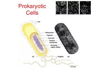
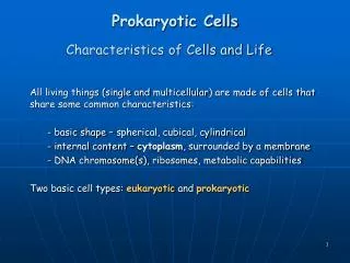
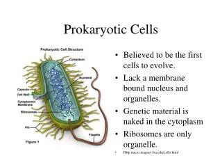
Microbiology Mrs. Hieneman. Anatomy and Physiology of Prokaryotic Cells. Bacterial Shape and Arrangement. Streptococcus chain. Sarcinae cube . Staphylococcus aureus cluster. Spiral-shaped bacterial cell. Prokaryotic Cell Structure. Cytoplasmic Membrane.

E N D
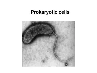
Microbiology Mrs. Hieneman Anatomy and Physiology of Prokaryotic Cells
Cytoplasmic Membrane • Surrounds cytoplasm and defines boundaries of cell • Acts as barrier, but also functions as an effective and highly discriminating conduit between cell and surroundings • Made up of phospholipid bilayer
Movement of Molecules through Cytoplasmic Membrane • Several ways for molecules to move through membrane • Simple Diffusion • Osmosis • Facilitated Diffusion • Active Transport
Simple Diffusion • Does not require expenditure of energy • Process by which some molecules move freely into and out of the cell • Small molecules such as carbon dioxide and oxygen
Figure 4.18: The principle of osmosis - Overview. Glass tube Rubber stopper Rubber band Sucrose molecule Cellophane sack Water molecule (a) At beginning of osmotic pressure experiment (b) At equilibrium Cytoplasm Solute Plasma membrane Cell wall Water (c) Isotonic solution — no net movement of water (d) Hypotonic solution — water moves into the cell and may cause the cell to burst if the wall is weak or damaged (osmotic lysis) (e) Hypertonic solution — water moves out of the cell, causing its cytoplasm to shrink (plasmolysis)
Transport Proteins • Transport proteins (or transporters) responsible for: • Facilitated Diffusion • Active Transport
Figure 4.17: Facilitated diffusion. Transported substance Transporter protein Outside Plasma membrane Inside Glucose
Cell Wall • Composed of peptidoglycan • Comprised of alternating NAG and NAM molecules • Attached to each NAM is four amino acid peptide: tetrapeptide
Categories of Bacteria • Two Major Categories: • Difference due to difference in chemical structures of their cell walls • Gram positive: stains purple • Gram negative: stains red
Gram + Cell Wall • Thick Layer of Peptidoglycan • Contains techoic acid: chains of ribitol-phosphate or glycerol-phosphate to which sugars or alanine attached • Techoic Acid sticks out above the peptidoglycan layer
Gram – Cell Wall • More complex than Gram + cell wall • Thin layer of peptidoglycan • Sandwiched between the cytoplasmic membrane and outer membrane • Outside of peptidoglycan is outer membrane
Outer Membrane • Unlike any other membrane in nature • A lipid bilayer with the outside layer made of lipopolysaccharides instead of phospholipids • Also called LPS • Contains Porins
Periplasm - Region between cytoplasmic membrane and the outer membrane - Gel-like fluid • Filled with secreted proteins and enzymes
External Structures • Glycocallyx • Flagella • Axial Filaments • Fimbrae and Pili
Glycocallyx • Gel-like structure • Functions in protection and attachment • Two types- capsule and slime layer • Involved in attachment, enabling bacteria to stick to teeth, rocks • Enables bacteria to brow as biofilm
Filamentous Protein Appendages • Anchored in membrane and protrude from surface • Flagella: long structure responsible for motility • Fimbrae and Pili: shorter, responsible for attachment
Four types of bacteria with flagella • Montrichious- one flagella • Amphitrichous- flagella at both ends • Lophitrichous- many flagella at the end of the cell • Peritrichous- flagella all over entire cell
Axial Filament • Present in Spirochetes • Attach at end of cell, spiral around, underneath an outer sheath • Move like a corkscrew
Fimbrae and Pili • Shorter and surround the cell • Similar structural theme to filament of flagella • Fimbrae- enable cell to adhere to surfaces, including other cells • Pili- join bacterial cells in preparation for the transfer of DNA from one cell to another
Cytoplasm • Substance of cell inside the cytoplasmic membrane • About 80% water • Thick, aqueous, semitransparent, elastic
Chromosome • Found within a central location known as nucleoid • Single, circular, double stranded • Consists of all DNA required by cell
Plasmids • Some bacteria contain plasmids- small circular double-stranded DNA • Typically cell does not require genetic information carried on plasmid • However, it may be advantageous
Ribosomes • Site of protein synthesis • Relative size and density of ribosomes and their subunits expressed as distinct unit (S) • Two units of prokaryotic ribosomes: 50S + 30S= 70S • Eukaryotic ribosomes: 80S
Figure 4.19: The prokaryotic ribosome. 50S 50S 30S 30S (a) Small subunit (b) Large subunit (c) (c) Complete 70S ribosome
Inclusions • Store excess nutrients • Examples: Polysaccharide granules- glycogen and starch • Lipid inclusions • Metachromatic granules- inorganic phosphate that can be used to synthesize ATP
Endospores • Occurs in members of genera Bacillus and Clostridium • Dormant cell produced by a process called Sporulation • Germination- when they exit the dormant state and then become a vegetative cell • Several species of endospore formers can cause disease