Understanding Bone and Cartilage Development: Growth Functions and Ossification Processes
This comprehensive overview explores the development and growth functions of bones and cartilage, highlighting their essential roles in support, movement, and protection. It details the types of skeletal cartilage—hyaline, elastic, and fibrocartilage—and the processes of ossification, including intramembranous and endochondral ossification. The text discusses bone histology, cell types, and the regulatory effects of hormones and vitamins on bone growth. Additionally, it covers the structure of bones, types of fractures, and the intricate bone repair process.

Understanding Bone and Cartilage Development: Growth Functions and Ossification Processes
E N D
Presentation Transcript
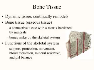
BONE TISSUE DEVELOPMENT and GROWTH
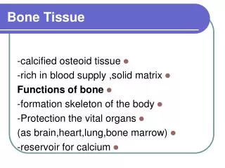
FUNCTIONS • Support/Movement • Protection • Mineral reservoir • Site of blood cell production • Storage of fat
Skeletal Cartilage • Consists primarily of water • Contains no nerves or blood vessels • Surrounded by perichondrium • Dense irregular connective tissue • Maintains shape
Skeletal Cartilage • Basic components • Chondrocytes in lacunae • Extracellular matrix with jellylike ground substance • 3 Types • Hyaline • Elastic • Fibrocartilage
Hyaline Cartilage • Most abundant • Fiber not detectable • Locations • Articular cartilage • Costal cartilage • Respiratory cartilage • Nasal cartilage
Elastic Cartilage • Contains more elastic fibers; more flexible • Found in ear and epiglottis
Fibrocartilage • Highly compressible • Great strength • Locations • Knee • Vertebral disks
Cartilage Growth • Two methods • Appositional – adds to outside • Interstitial – growth from inside • Growth stops during adolescence
Bone Histology - Cell Types • Osteocytes – mature bone cells • Osteoblasts – bone forming cells • Osteoclasts – bone destroyers
BONE OSSIFICATION Process by which tissue becomes bone Also called osteogenesis
Bone Formation • Bone formation begins approx. 8 weeks into fetal development from a skeleton that is mostly fibrous membranes and cartilage • Intramembranous ossification – bone forms from the fibrous membranes • Endochondral ossification – bone forms from hyaline cartilage
Intramembranous Ossification • Osteoprogenitor (mesenchymal) cells in fibrous C.T. develop into osteoblasts • Osteoblasts secrete collagen matrix • Calcification occurs in ossification centers; forming a network of bone rather than layers • Bony plates form which are later converted into compact bone • Flat bones only; skull & clavicles • Fontanelles are areas not ossified at birth
ENDOCHONDRAL OSSIFICATION • Forms most bones • Hyaline cartilage model in shape of the bone initially; a pH change causes cartilage to calcify and the cells to die • Primary ossification center forms as blood vessels from periosteum and osteoblasts invade calcified cartilage • Matrix formed (osteoid= unmineralized bone matrix) • Ossification occurs = calcium salts deposited • Primary centers form before birth; Secondary centers form 8th month dev.
Epiphyseal Plate • Cartilage region between primary and secondary ossification centers • Responsible for postnatal bone growth • Zone of resting cartilage • Growth Zone – mitosis occurs • Transformation Zone – cartilage matrix deteriorates • Osteogenic Zone - bone salts deposited
Calcium regulation • Calcium is most abundant mineral in the body; 99% located in the bone • Regulated by two hormones: PTH (parathyroid hormone) and calcitonin • PTH - raises blood calcium levels • Calcitonin - lowers blood calcium levels
Hormones and Vitamin Effect on Bone Growth • Testosterone • Estrogen • Growth Hormone • Throxine - • Vitamin D – calcium absorption
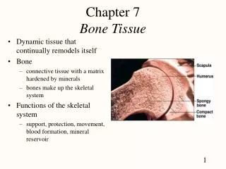
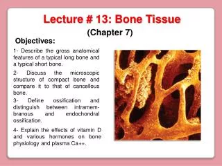
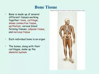
Compact Bone also called dense bone hard, strong and solid bone that forms the outer layer of all bone provides support, protection and resists stress Contains osteons Cancellous also called spongy bone found more toward the inner portion of bone open lattice-work of struts and plates that serves to store bone marrow Trabeculae Bone Types
Haversian canal Volkmann’s canals Lamellae Lacunae Canaliculi Osteons = Haversian System
Bone Types • Long – arms and legs • Short – wrist and ankle • Sesamoid – forms within a tendon (patella) • Flat – sternum, scapula, ribs, skull • Irregular – vertebrae & coxal bones
Structure of a Long Bone • Diaphysis • Epiphysis • Articular cartilage • Periosteum – connective tissue covering bone • Medullary cavity • Endosteum – connective tissue; lines inside
Bone Fractures • Open ( Compound) – penetrates skin • Closed (Simple) • Partial/Complete - Greenstick • Comminuted – broken into 3 or more pieces
Bone Repair • Formation of clot ( hematoma) • Callus ( soft followed by hard) • Mineralization of callus by calcium & phosphorus • Remodeling