Bone Tissue
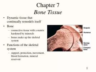
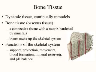
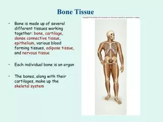
Bone Tissue. -calcified osteoid tissue -rich in blood supply ,solid matrix Functions of bone -formation skeleton of the body -Protection the vital organs (as brain,heart,lung,bone marrow) -reservoir for calcium. Shape: -Long bone:(as limbs), -short bone :(as hand,foot)

Bone Tissue
E N D
Presentation Transcript
Bone Tissue • -calcified osteoid tissue • -rich in blood supply ,solid matrix • Functions of bone • -formation skeleton of the body • -Protection the vital organs • (as brain,heart,lung,bone marrow) • -reservoir for calcium
Shape: • -Long bone:(as limbs), -short bone :(as hand,foot) • -Irregular bone:(as vertebrae) • -Flat bone:(as skull,scapula,sternum,iliac bones,ribs
Type of Bone • 1-compact solid bone : • present in shafts of long bone&in the outer thin layer of the spongy bone in old age • 2-spongy bone: • present in epiphses of long bones,ribs vertebrae • Flat bones as skull,scapula,sternum&sacrum
Histological slides taken from compact • bone can prepared by two methods • 1-Decalcified compact bone: treated with nitric acid • 2-Ground compact bone: dry bone is ground to demonstrate haversian canals,volkmans canals, • lacunae&canaliculi
Structures of bone • Bone is formed of • 1-bone matrix: formed of calcified lamellar of type I collagen • 2-bone cells: • osteogenic cells,osteoblasts,osteocytes&osteoclasts • 3-periosteum:covering layer of bone from outside • 4-Endosteum: lining layer of bone from inside
Chemical formation of bone matrix • 1-organic substance(35%)formed of protein&carbohydrate • 2-Inorganic substance(65%) formed of minerals as calcium phosphate
Types of bone cells • There are four types of bone cells • 1-osteogenic cells • 2-osteoblasts cells • 3-osteocytes cells • 4-osteoclast cells
1-Osteogenic Cells(osteoprogenerator cells) • develop from embryonic mesenchymal cells or from pericytes cells • -Present: periosteum,endosteum&bone marrow cavities • -spindle shaped cells with pale cytoplasm&flat nuclei • -can divide during growth of bone during healing of fractured bone
2-osteoblast Cells • Origin from osteogenic cells • Oval cells with eccentric rounded nuclei • The cell membrane has few cytoplasmic process • The cytoplasm is deep basophilic around the nucleus
there is an unstained area ( golgi apparatus) • The cytoplasm is rich in RNA, • endoplasmic reticulum&golgi body • Osteoblast cannt divid,they are bone building cells
Site: • osteoblast are found periosteum,endosteum&in bone marrow cavities
Function • 1- synthesise of protein • 2-osteoblasts secrete matrix vesicle rich in enzymes • a-Alk Ph. Enzymes(facilitate deposition of calcium) • b-pyrophosphatase enzymes(retard process of calcification) • 3-osteoblast change into osteocytes surround by lacunae&by calcified matrix
3-osteocytes • -Mature non-dividing bone cell • -Present inside lacunae • -Cytoplasm is basophilic • -Rich in phosphatase enzymes • -Nucleus is oval¢ral
Origin: • osteocytes are considered as mature osteoblast surround by calcified matrix • Function: • 1-they form the bone matrix vesicle rich in enzymes • 2-they release calcium from bone to the circulating blood
4-Osteoclast Cells • Origin:from the circulating blood monocytes large cell with irregular cell membrane • Each cell contain from 4-50 nuclei • Site:they are present in bone marrow cavities &endosteum of bone
Functions of osteoclast • 1-Concerned in bone resorption during ossification • 2- Secrete enzymes during ossification • 3-Secrete acids which play arole in decalcification of bone matrix • 4-Remove bone debris during ossification
Microscopic structures of compact bone • -transvers section in the shaft of adult long bone • 1-Haversian system • 2-Interstitial lamellae • 3-Outer&inner circumferential lamellae under periosteum&endosteum • 4-Periosteum:cover the bone • 5-Endosteum:line the bone
COMPACT BONE Volkmann’s Haversian canal canal Ground Bone outer Volkmann’s lamellae canal circumferential Haversian systems (osteons) inner circumferential lamellae spongey bone interstitial Haversian system lamellae Haversian canal
Periosteum • -avascular C.T.membrane • -formed of two layers • a-Outer fibrous layer:formed of collagenous fibers,fibroblast&fibrocytes cells,nerve,blood vessls • b-Inner osteogenic layer:formed of osteogenic spindle cells
PERIOSTEUM Sharpey’s Fibers (regions of tendon attachment) Periosteum Decalcified Tibia (transverse section) bone compact compact bone endosteum marrow periosteum osteoblasts
Function of periosteum • 1-provides an attachment for muscles,tendon&ligments • 2-provides the bone with blood supply&nourishment • 3-provides formation bone during growth&after fracture
The Endosteum • It formed of avascular C.T.membrane rich in osteogenic cell,osteoblast&osteoclast cells • Function of endosteum • 1-supplies bone with blood supply&nutrition • 2-Its osteogenic cells,osteoblast cells&osteoclast are concerned with bone formation during growth
Volkmans Canals • These are transvers or oblique canals,they connect the haversian canals together • -connect the haversian canals with the periosteum or endosteium • -they are lined by endosteum&contain blood vessel
Haversian System or osteon • Its formed of • A-Haversian canal contain C.T.&blood vessels&osteogenic cells • B-concentric bone lamellae:formed of • 4-20 layers of bone lamellae • C-Osteocytes these are the mature bone cells present inside their lacunae
The external circumferential lamellae • Formed of calcified osteoid tissue in which osteocytes are embedded present under the periosteum&arranged parallel
The internal circumferential lamellae • These lamellae are formed of calcified osteoid tissue present adjacent to the lamellae
The interstitial or the interhaversian lamellae • These are formed of calcified osteoid tissue • Present between the haversian system • Osteocytes in these interstitisl lamellae are irregular arranged
Spongy or cancellous bone • Long&short bone are formed externally of compact bone • Endosteum are irregular due to presence of spongy bone • Site: • Vertebrae,ribs,flat bones.epiphyses of long bones
Grossly Areas with numerous interconnecting cavities called spongy ( cancellous ) bone • In long bone : -Epiphyses : spongy bone covered by a thin layer of compact bone -Diaphysis : compact bone with small spongy bone on it,s inner surface around the bone marrow • Short bones : a core of spongy bone completely surrounded by compact bone • The flat bone : have two layers of compact bone called plates separated by a layer of spongy bone called the diploë
Spongy bone periosteum woven matrix osteocytes osteoblasts osteoblasts bony spicules osteocytes Ground Substance (glycosaminoglycans) Collagen (reinforcing) Bone Mineral (calcium phosphate) osteocytes
Ossification • Ossification : process formation of bone leads to growth • Methods for bone ossification • 1-Intramembranous ossification(occurs in mesenchymal membranes) • 2-Intracartilagenous (occurs in cartilage models)
Mechanism of ossification • Two processes • A.Bone formation • B.Bone resorption
1-Intramembranous ossification • It occurs in flat bones of the face,skull&clavicle • Site of feature bone is occupied by mesenchymal membrane formed of matrix blood capillaries&mesenchymal cells,this membrane transformed into spongy bone by the following steps
1-Acentre of ossification appears in middle of mesenchymal membrane,blood supply increased&the mesenchymal transformed into osteogenic cells……transformed to osteoblasts
2-The newly osteoblasts,synthesis the organic component of the matrix rich in ph. Enzymes which can deposit ca++ • Osteoblast surrounded by solid matrix then transformed into osteocytes • 3-the trabeculae of the newly formed spongy bone
4-the osteogenic cells formed periosteum&endosteum of spongy bone • 5-growth&remodlling of bone occurs by deposition of new bone by the osteoblast&resorption of irregular bone by osteoblasts
INTRAMEMBRANOUS OSSIFICATION Mesenchymal Cells (connected by processes) Collagen and Matrix Calcified Bony Primary Matrix (trabeculae) Spongiosa Osteoblasts Osteoclasts Osteocytes osteocytes posteoprogentor cells osteoblasts Intramembranous Ossification monocytes osteoclasts Skull- frontal, parietal, occipital temporal bones mandible- jaw
2-Intracartilagenous ossification • :Occurs in the long bones which were originally formed of hyaline cartilage replaced by bone ossification • Start as primary center of ossification in the middle part of the long bone(Diaphysis) • Secondary center appears end of long bones(Epiphysis
Stages of Intracartilagenous Ossification • 1-Resting satge of hyline cartilage • Present in the region of the growing zone of the long bone • 2-proliferative stage of cartilage cells • Increase in number of young cartlage cells,chondrocytes divid give small chondrocytes arranged in rows
3-Maturation of cartilage cells • Small chondrocytes increase in size&become mature cells • 4-calcification sage of cartilage • ALK.Phosph. enzymes in the mature cartilage cells deposite calcium phosphate&carbonate in the matrix
At the same time some of osteogenic cells under the perichondrium changes into osteoblast which can form alayer of calcified tissue around the perichondrium forming periosteal collar
5-Stage of invasion • Dead cartilage cells are replaced by blood capillaries,osteogenic cells,blood monocytes&mesenchymal cells,these invading cells can be changed into osteoblast,osteoclasts&bone marrow cells
6-Satges of spongy bone formation • These osteoblasts start to form the trabculae of spongy bone by depositing calcium to form the calcified trabeculae of spongy bone
7-Satge of internal reconstraction bone • Spongy bone is transformed into compact bone,some blood monocytes change in osteoclasts • Osteoclast will destroy the central irregularities of spongy bone forming regular endosteum
8- Satge of complete ossification • Haversian systems are formed as follows • Osteoblast will arrange themselves,concentrically around B.V. taking from them ca++&nutrition
-osteoblasts can form calcified bone lamellae around the central B.V.this formed longitudinal canal around blood vessels is called Haversian canal • The processes goes on several times until mature Haversian system are formed
HAVERSIAN SYSTEMS Ground Bone lamella canaliculae Haversian canal (artery & vein or capillary) osteocyte canaliculae lacuna