Download
1 / 59
800 likes | 1.91k Views
Traumatic Brain Injury (TBI). Adult Health II Traumatic Brain Injury—Part 2. Jerry Carley RN, MA, MSN, CNE Summer 2010. Concept Map: Selected Topics in Neurological Nursing. PATHOPHYSIOLOGY Traumatic Brain Injury Spinal Cord Injury Specific Disease Entities :

E N D
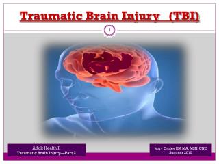
Traumatic Brain Injury (TBI) Adult Health II Traumatic Brain Injury—Part 2 Jerry Carley RN, MA, MSN, CNE Summer 2010
Concept Map: Selected Topics in Neurological Nursing PATHOPHYSIOLOGY Traumatic Brain Injury Spinal Cord Injury Specific Disease Entities: Amyotropic Lateral Sclerosis Multiple Sclerosis Huntington’s Disease Alzheimer’s Disease Huntington’s Disease Myasthenia Gravis Guillian-Barre’ Syndrome Meningitis Parkinson’s Disease ASSESSMENT Physical Assessment Inspection Palpation Percussion Auscultation ICP Monitoring “Neuro Checks” Lab Monitoring PHARMACOLOGY --Decrease ICP --Disease Specific Meds Care Planning Plan for client adl’s, Monitoring, med admin., Patient education, more…based On Nursing Process: A_D_P_I_E Nursing Interventions & Evaluation Execute the care plan, evaluate for Efficacy, revise as necessary
Objectives • Recall anatomy and physiology of the brain & cranial nerves • Explain pathophysiology of various brain (head) injuries • Detail signs, symptoms and prevention of Increased Intracranial Pressure (ICP) • Demonstrate effective use of Glasgow Coma Scale • Discuss medical & nursing management of brain injuries
Prevent Secondary Injury !!! Meaningful recovery of function after head injury is possible IF secondary injuries are prevented or minimized
Secondary Brain Injury • Any physiological event that can occur within minutes, hours, or days after the initial injury and leads to further damage of nervous tissue • Secondary Injury is mostly due to Increased ICPcaused by hypotension, hypoxia, intracranial bleeding, seizures
Brain Injury Management Frequent Re-assessments + Rapid Response
Be Vigilant for Increased ICP ! To understand intracranial pressure, think of the skull as a rigid box. After brain injury, the skull may become overfilled with swollen brain tissue, blood, or CSF. The skull will not stretch like skin to deal with these changes. The skull may become too full and increase the pressure on the brain tissue. This is called increased intracranial pressure. Foramen Magnum ICP Peaks 48 – 72 hours after injury
Monitor: Neuro Checks q 15 minutes • Vital Signs Q15 minutes • Glasgow Coma Score Q15 minutes
Expanded Neuro Assessment Tool
EARLY Signs of ↑ ICP • Slight LOC changes ***MOST IMPORTANT**** • 2. Pupils sluggish / Impaired eye movement • 3. Limb strength changes • 4. Headache
Change in • Level Of Consciousness (LOC) • ***MOST IMPORTANT**** • + • EARLIEST • Indicator of neurological deterioration
Cushing’s Triad: Signs of ↑ ICP • Blood Pressure • Systolic BP Increases • Diastolic BP Decreases • Pulse Decreases Widening Pulse Pressure Bradycardia *** You will also see listed in some resources: --Irregular Respirations (Cheyne-Stokes) --Elevated Temperature (Hyperpyrexia)
TREND Re-Assessment Data + COMPARE to Baseline Assessment Data Temp Pulse BP
LATE(R) Signs of ↑ ICP • Further decreased LOC • Cushing’s Triad / Reflex • Abnormal respiration patterns • Pupils asymmetrical / Dilated • Projectile vomiting • Hemiplegia / decorticate or decerebrate posturing
Brain Herniation occurs when a part of the brain pushes downward inside the skull through the opening that leads into the neck (Foramen Magnum)
Tentorial (Brain) Herniation Normal
ABI Nursing Interventions • Continuous monitoring of Vitals, PERL and Glasgow Coma Score • Report client condition changes ASAP • Maintain airway patency (eg positioning, suctioning, etc) • Minimize cerebral edema • Maximize cerebral perfusion • Implement seizure precautions / Siderails • Provide emotional support • Address all self-care deficits
Neurosurgeon drilling prior to placing an intracranial pressure monitor
Normal ICP for adults: 10 to 15 mm Hg
ABI Priority Nursing GOALS * Minimize cerebral edema * Maximize cerebral perfusion
ABI Nursing Interventions • Continuous monitoring of Vitals, PERL and Glasgow Coma Score • Report client condition changes ASAP • Maintain airway patency BUT… • Avoid suctioning or Hyperventilate with 100% O2 FIRST
ABI Nursing Interventions • Implement seizure precautions / Siderails • Phenytoin (Dilantin) (prevent / treat Sz) • Maintain head midline (neutral position) • HOB > 30 degrees
ABI Nursing Interventions • Address all self-care deficits…BUT • Avoid clustering activities • Provide emotional support
ABI Nursing Interventions • High dose barbituates > induced coma *decreases metabolic demands* • Pharmacological paralysis • Avoid overstimulation: - Dark quiet room - Limit visitors appropriately - Speak softly - Limit dialogue – keep topics light hearted
Minimize Cerebral Edema • Mannitol (Osmitrol) + Urinary catheter • Fluid restriction (I & O)…? • Dexamethasone / Decadron(Know side effects!) • Prevent / Treat fever • Prevent Infections (closed STERILE monitoring system)
Minimize Cerebral Edema Maintain Cerebral perfusion pressure MAP of 50 – 70 mm Hg Prevents Hypoxia (Hypercarbia)
IfBPtoo low…then O2perfusion is poor…andBrain Can’t Function
Optimize Cerebral Perfusion • Keep head position midline • HOB elevated ( 30 - 60 degrees ) • Oxygen **** • Sedate prior to activity • Minimal ADL movement of client
Teach Client / Family • Minimal stimulation environment • No coughing, no straining, no hard laughing • Head midline + Bedrest + HOB elevated • S & S to report to nurse ASAP (Headache, drainage, etc) • Purpose + frequency of neuro checks • Medication regime (Narcotics, diuretics, stool softeners, etc) • Medical interventions (Tests, traction, logrolling, surgery, etc)
Cerebral Concussion • A ‘concussion’ is a relatively mild form of traumatic brain injury that results in temporaryneurological changes • No apparent structural damage • Usually involves unconsciousness for a few seconds or minutes • Frontal lobe = bizarre irrational behavior • Temporal lobe = amnesia or disorientation
Discharge …. • Mild concussion & neurological stability = usually will not require hospital admission • However !!! Must be observed by a reliable companion for at least 12 hours • No alcohol for several days • No pain medications stronger than Tylenol
Cerebral Contusion • More severe • Brain bruised • Possible surface hemorrhage • Initially appears like shock • Can have B & B incontinence • Can be aroused…briefly
IntraCerebralHemorrhage IntraCranial Hemorrhage Bleeding within the tissue of the brain Bleeding within the cranial vault
IntraCranialHemorrhage Bleeding within the cranial vault
IntracranialEpidural / Extradural Hematoma - Between skull and dura - Extreme emergency - Mostly arterial
Subdural Hematoma Between dura and brain Mostly venous
Subdural Hematoma 3 Types: Acute Sx in 24 – 48 hours Subacute Sx in 48 hours – 2 weeks Chronic Sx in 3 weeks – months Common in elderly after even minor injury Often misdiagnosed as stroke
Head trauma leading to subdural hematoma and intracranial hypertension
Subarachnoid Hemorrhage • Subarachnoid space is brain surface where blood vessels that supply the brain are located • Common causes of subarachnoid hemorrhage are trauma to “Circle of Willis” aneurysms and congenital arteriovenous malformations (AVM) • Unique S & Ss: - Sudden & unusually severe headache & loss of consciousness - Neck pain & ridigity (nuchal rigidity) d/t meningeal irritation • Untreated, the blood supply to a given area of the brain may fall so low that the brain tissue dies resulting in a stroke
IntraCerebralHemorrhage Bleeding within the tissue of the brain
IntracerebralHemorrhage / Hematoma Causes: - Force is exerted to the head over a small area (missile injuries, bullet wounds, etc) - Systemic hypertension causes degeneration and rupture of blood vessels - Tumors - Bleeding disorders