The basic neurological examination
The basic neurological examination. Dr. Szathmári Miklós Semmelweis University First Department of Medicine 12. Dec. 2011. The neurological examination. Mastery of the complete neurological examination is usually important only for physicians in neurology

The basic neurological examination
E N D
Presentation Transcript
The basic neurological examination Dr. Szathmári Miklós Semmelweis University First Department of Medicine 12. Dec. 2011.
The neurological examination • Mastery of the complete neurological examination is usually important only for physicians in neurology • However, knowledge of the basics of the examination, especially those component that are effective in screening for neurological dysfunction is essential for general physicians • A screening examination done in the suggested way lasts 3-5 min.
Mental status examination • During the interview, look for difficulties with communication and determine whether the patient has recall and insight into recent and past events. • If the history raises any concern for abnormalities of higher cortical function or if cognitive problems are observed during the interview, then detailed testing of the mental status is indicated.
Elements of mental status examination • Level of consciousness • The patient’s relative state of awareness of the self and the environment (ranged: fully awake-comatose). • The doctor has to describe the responses to the minimum stimulus necessery to elicit a reaction. • Loss of oculocephalic reflex (doll’s eye movements) in a comatose patient suggest a lesion of midbrain or pons (Holding open the upper eyelids turn the head quickly, first to one side and than to the other. Normally the eyes move in the opposite direction as if still gazing ahead in their initial position). • Orientation • Is tested by asking to state his or her name, location and time
Level of consciousness • Is the patients awake and alert? • Does the patient seem to understand your questions and respond appropriately or is there a tendency to lose track of the topic and fall silent or even sleep? If the patient does not respond, escalate the stimulus in steps: • Speak to the patient in a loud voice • Shake the patients gently • Brief painful stimulus such as squeeze of the trapezius muscle) • If there is no response that means severe reductions in the level of consciousness (stupor or coma)
Elements of mental status examination • Speech • Articulation, rate, rhythm, etc. • Memory • Immediate memory: saying a list of three items, and the patient repeat the list immediately • Short-term memory: to recall the same items 10 min later • Long-term memory: how well the patient is able to provide a coherent chronologic history of his or her personal events • Abstract thought • Can be tested by asking the patient to list items having the same attributes (e.g. a list of four-legged animals) • Calculation ability • Serial subtraction 7 from 100 or 3 from 20
Cranial nerve examination 1. • CN I.(olfactory) – Testing is usually omitted unless there is suspicion for inferior frontal lobe disease (e.g. meningeoma) • With eyes closed, ask the patient to sniff a mild stimulus such as toothpaste or coffee and identify odorant. • Symptom:hyposmia, anosmia • CN II.(optic)- Checking visual acutity and testing the visual fields by confrontation, by comparing the patient’s visual fields to your own. • Face the patient at a distance of appr. 0.6-1.0 m and place your hands at the periphery of your visual fields in the plane that is equidistant between you and the patient. Instruct the patient to look directly at the center of your face and to indicate when and where he or she sees one of your finger moving. • CN III, IV, VI (oculomotor, trochlear, abducens) Describe the size and shape of pupils and reaction to light and accomodation, check extraocular movement • The patients’s eyes converge while following your finger as it moves toward the bridge of nose • Ask the patient to keep his or her head still while tracking the movement of the tip of your finger • CN V (trigeminal) – Examine sensation within the three territories of the branches of the trigeminal nerve on each side of the face • Light touch and temperature • Other modalities : corneal reflex and jaw clench is indicated when suggested by the history)
Cranial nerve examination 2. • CN VII (facial) - look for facial assymetry at rest and with spontaneous movement • Eyebrow elevation, forehead wrinkling, eye closure, smiling and cheek puff • Weakness of the lower two-third of the face with preservation of upper third suggest an upper motor neuron lesion • Weakness of an entire side suggest a lower motor neuron lesion. • CN VIII (vestibulocochlear) – Check the patient’s ability to hear a finger rub or whispered voice with each ear • Further testing for air versus mastoid bone conduction etc. Should be done if an abnormality is detected by history or examination. • CN IX, X (glossopharyngeal, vagus) – Observe the position and symmetry of palate and uvula at rest and with phonation („aah”) • Symptoms of injury: paresis of the palate, nasal voice, disturbance with swallow, hoarseness. • CN XI (spinal accessory) • Chek shoulder shrug (trapezius muscle) and head rotation to each side (sternocleidomastoid) against resistance • CN XII (hypoglossal) • Inspect the tongue (In case of the unilateral paresis the tongue deviates in the direction of the normal side)for atrophy or fasciculation, position, and strength when extended against the inner surface of the cheeks on each side
Cranial nerve examination 3. • The bare minimum: check • the visual fields • the pupil size • the pupil reactivity • the extraocular movements • the facial movements • Bulbar paralysis: lesion of the IX-XII. cranial nerves: disturbance of swallowing, breathing and dysarthria, and atrophy of the tongue. • Pseudobulbar paralysis: the same except of the tongue atrophy.
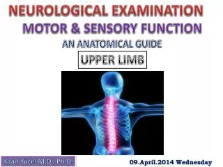
Motor examination 1. • Observation of muscle • Appearance • Inspection and palpation of muscle group with patient in a comfortable and symmetric position • Check atrophy, hypertrophy and tenderness • Involuntary movements • present at rest: myoclonus, tics • during maintained posture: pill-rolling tremor of Parkinson’s disease • with voluntary movements: intention tremor of cerebellar disease, etc. • Tone is tested by measuring the resistance to passive movement of relaxed limb. • While the patient is supine the examiner’s hand are placed behind the knees and rapidly raised • With normal tone the ankles are drag along the table surface for a variable distance before rising • Increased tone results in an immediate lift of the heel off the surface • Cogwheel rigidity: in which passive motion elicits jerky interruptions in resistance, is seem in parkinsonism
Motor examination 2. • Observation of the muscle strength • Pronator drift method: the patient is asked to hold both arms fully extended and parallel to the ground with eyes closed. This position should be maintained for 20-30 sec. • Any flexion at the elbow or fingers or pronation of the forearm, especially if asymmetric, is a sign of potential weakness. • Examination of active movements • Example: test flexion at the hip: by placing your hand on the patient’s thigh and asking the patient to raise the leg against it • Paralysis: no movement • Severe weakness: movement with gravity eliminated • Moderate weakness: movement against gravity but not against mild resistance • Mild weakness: movement against moderate resistance • Full strendth
Motor examination 3. • Muscle stretch reflexes • Biceps (C5,6), triceps (C7,C8), patellar (L3,4), and Achilles (S1,S2) • The patient should be relaxed and the muscle positioned midway between full contraction and extension • Reflexes may be enhanced by asking the patient to voluntarily contract other, distant muscle group (JENDRASSIK’S MANEUVER)
Jendrassik’s maneuver If the patient’s reflexes are symmetrically diminished or absent, it should be reinforced by a technique involving isometric contraction of other muscles that may increase reflex activity. If the knee reflexes are diminished, reinforce them by asking the patient to lock fingers and pull one hand against the other.
Cutaneous reflexes • The plantar response: With a key or the wooden end of an applicator stick, stroke the lateral aspect of the sole from the heel to the ball of the foot, curving medially across the ball: • The movement of the toes normally flexion • Dorsalflexion of the big toe often accompanied fanning of the other toes – Babinski response indicates upper neuron disease. • Superficial abdominal reflexes • Gently stroking the abdominal surface near to umbilicus in a diagonal fashion with a sharp object • Normally the umbilicus will pull toward the stimulated quadrant • Absent of abdominal reflex indicates upper motor neuron lesions • Cremasteric reflex • Ipsilateral elevation of testicle following stroking of the medial thigh (mediated by L1 and L2)
Sensory examination • Pain and temperature, impulses for which are carried in the spinothalamic tracts of the cord • Position and vibration, impulses for which are carried in the posterior columns • Light touch, impulses for which are carried along both of these pathways • Discriminative sensations, which depend on some of the above sensations but also require cortical judgments
Screening of sensory system • Pain sensation in the hand and the feet • It is tested using a sharp safety pin • Anelgesia-hypalgesia-hyperalgesia • Light touch • It is assessed with single, very gentle touch of the examiner’s finger • Anesthesia – absence of touch sensation, hypesthesia-decresed sensitivity, hyperesthesia- increased sensitivity • Vibration • It is tested using 128 Hz tuning fork applied to the distal pahalanx of the great toe • Temperature • It is assessed using a metal object that has been immersed in a cold and warm water • Disciriminative sensation • Such as graphesthesia –with a blunt end of a pen, draw a large number in the patient’s palm
Special maneuvers • Meningeal sign (in case of suspition of inflammation of the meninges or bleeding (subarachnoid) • The patient is supine position. • Place your hand behind the patient’s head and flex the neck forward, until the chin touches the chest. Note resistance or pain. Watch also for flexion of the patient’s hips and knees during the maneuver – The name of the positive response is Brudinski’s sign. • Kernig’ sign: Flex one of the patient’s legs at both hip and knee, then straighten the knee. Unusual resistance or pain – mostly when bilateral – suggests meningeal inflammation
Coordination examination • Coordination refers to the orchestration and fluidity of movements. • Part of this integration relies on normal function of the cerebellar and basal ganglia, however, coordination requires intact muscle strength and proprioceptive information • The minimum testing of cerebellar function: • Finger-to-nose test: the patients is asked to touch his or her index finger repetitively to the nose and then to the examiner’s outstreched finger, which moves with each repetition. • Heel-knee-shin maneuver: in the supine position the patient is asked to slide the heel of each foot from the knee down the shin of the other leg. • The accuracy, speed, and rhythm are observed.
Gait examination • Normal gait requires multiple systems – including strength, sensation, and coordination – function in a highly integrated fashion. • The patient should be observed while walking and turning normally, walking on the heels, on the toes, and walking heel to-toe along a straight line. • Decreased arm swing on one side – corticospinal tract disease • Short-stepped gait – parkinsonism • Broad-based unstable gait – ataxia (may be due to cerebellar disease, loss of position sense, or intoxication) • High-stepped, slapping gait – posterior column or peripheral nerve disease
Localization of damage according to signs Harrison’s: Principles of Internal Medicine. 17th Edition. McGraw Hill (modified table)