Download

1 / 54
540 likes | 743 Views
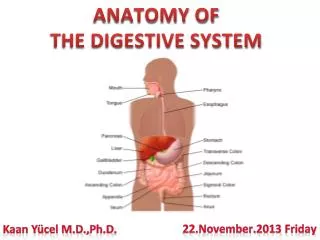
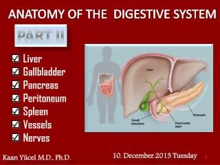
Yeditepe University Medical School Department of Anatomy. ANATOMY OF THE DIGESTIVE SYSTEM PART 1. 2.December.2011 Friday. Kaan Yücel M.D.,Ph.D. Oral Region. The oral region includes the oral cavity , teeth , gingivae , tongue , palate , and the region of the palatine tonsils .

E N D
Yeditepe University Medical School Department of Anatomy ANATOMY OF THE DIGESTIVE SYSTEM PART 1 • 2.December.2011 Friday Kaan Yücel M.D.,Ph.D.
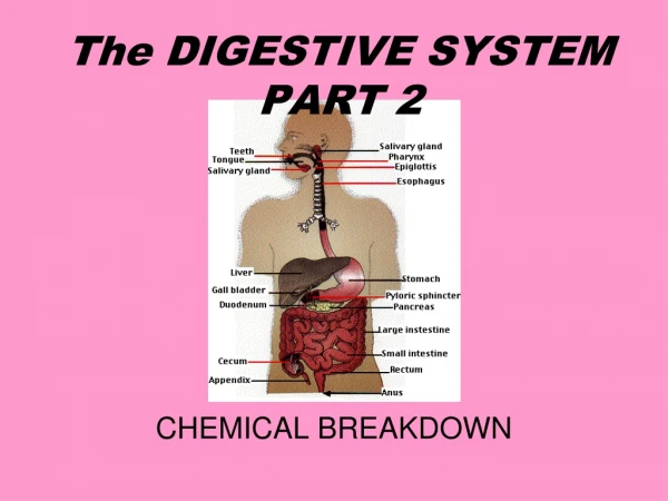
Oral Region The oral regionincludes the oral cavity,teeth, gingivae, tongue, palate, and the region of the palatine tonsils. The digestion starts here in the oral cavity. It is the place where the food is ingested and prepared for digestion in the stomach and small intestine.
Food is chewed by the teeth, and saliva from the salivary glands facilitates the formation of a manageable food bolus(L. lump). Deglutition (swallowing) is voluntarily initiated in the oral cavity. The voluntary phase of the process pushes the bolus from the oral cavity into the pharynx, the expanded part of the alimentary (digestive) system, where the involuntary (automatic) phase of swallowing occurs.
Oral Cavity (Mouth) Inferiortothenasalcavities . Extendsfromthelipstothepharynx.
Oral Cavity (Mouth) Has a roofandfloor, andlateralwalls. Opensontothefacethroughthe oral fissure. Continuouswiththecavity of thepharynx at theoropharyngealisthmus.
The oral cavity has multiplefunctions: Inletforthedigestivesysteminvolvedwiththeinitialprocessing of food, which is aidedbysecretionsfromsalivaryglands. Manipulatessoundsproducedbythelarynx. Can be usedforbreathingbecause it opensintothepharynx, which is a commonpathwayforfoodandair. Forthisreason, the oral cavity can be usedbyphysicianstoaccessthelowerairway.
Oral cavity (mouth) consists of two parts: Oral vestibule Oral cavity proper
Slit-likespacebetweentheteethandgingivae (gums) internallyandthelipsandcheeksexternally. • Enclosedbydentalarches. • Communicateswiththeexteriorthroughtheoral fissure(opening). Oral vestibule
Thelateralwall of thevestibule is formedbythecheek, which is madeupbythebuccinatormuscle. Thetone of thebuccinatormuscleandthat of themuscles of thelipskeepthewalls of thevestibule in contactwithoneanother.
Theduct of theparotidsalivarygland (Stensen’sduct) opens on a smallpapillaintothevestibuleoppositetheuppersecondmolartooth.
Thespacebetweentheupperandthelowerdentalarches. • Has a roofand a floor. • Theroof of themouth is formedbythe hard palate in frontandthesoftpalatebehind. • Oral cavityproper
Thefloor is formed • largelybytheanterior 2/3 of thetongue • and • bythereflection of themucousmembranefromthesides of thetonguetothegum of themandible.
Thesubmandibularduct of thesubmandibulargland (Warton’sduct)opensontothefloor of themouth on thesummit of a smallpapilla on eitherside of thefrenulum of thetongue.
Lips • Mobile, musculofibrous folds surrounding the mouth • Coveredexternally by skin and internally by mucous membrane. • Function as the valves of the oral fissure, containing the sphincter (orbicularis oris) that controls entry and exit from the mouth and upper alimentary and respiratory tracts. • .
Formthe movable walls of the oral cavity. The prominence of the cheek occurs at the junction of the zygomatic and buccal regions. • Cheeks (Buccae) External aspect- Buccal region Anteriorly by lips and chin Superiorly by zygomatic region Posteriorly parotid region Inferiorly by inferior border of mandible
Teeth • Thechieffunctions of theteethareto: • Incise, reduce, andmixfoodmaterialwithsalivaduringmastication. • Helpsustainthemselves in thetoothsocketsbyassistingthedevelopmentandprotection of thetissuesthatsupportthem. • Participate in articulation (distinctconnectedspeech).
The teeth are set in the tooth sockets. There are 20 deciduous teeth and 32 permanent teeth: four incisors, two canines, four premolars, and six molars in each jaw.
Gingivae (Gums) • Composed of fibroustissuecoveredwithmucousmembrane. • The gingiva proper (attached gingiva) is firmly attached to the alveolar processes of the mandible and maxilla and the necks of the teeth.
Formspart of thefloor of the oral cavityandpart of theanteriorwall of theoropharynx. • Tongue A mass of striatedmusclecoveredthmucousmembrane
Itsanteriorpart is in the oral cavityand is somewhattriangular in shapewith a bluntapex of tongue. The apex is directed anteriorly . Theroot of tongue is attachedtothemandibleandthehyoid bone.
Papillae Thesuperiorsurface of the oral part of thetongue is coveredbyhundreds of papillae. 4 types of papillae in thetongue:
The superior surface of the oral part of the tongue is covered by hundreds of papillae. 4 types of papillae in the tongue. The papillae in general increase the area of contact between the surface of the tongue and the contents of the oral cavity.
Muscles of theTongue IntrinsicandExtrinsicMuscles Intrinsic muscles: confined to the tongue, are not attached to bone. Extrinsic muscles: attached to bones and the soft palate.
Innervation of the tongue is complex and involves a number of nerves. Trigeminal nerve: sensation from 2/3 anterior tongue Glossopharyngeal nerve: sensation from 1/3 posterior tongue Taste from oral part: by the facial nerve Taste from pharyngeal part: by the glossopharyngeal nerve
Movements of theTongue Protrusion: Retraction: Depression: Shape changes:Intrinsic muscles
Movements of theTongue Depression:Hyoglossusmuscles on bothsidesactingtogether Retractionandelevation of theposteriorthird:Styloglossusandpalatoglossusmuscles on bothsidesactingtogether Shapechanges:Intrinsicmuscles
Palate • Formsthearchedroof of themouthandthefloor of thenasalcavities. • Separatesthe oral cavityfromthenasalcavitiesandthenasopharynx, thepart of thepharynxsuperiortothesoftpalate. • Consists of 2 regions: • Hard palateanteriorly • Ssoftpalateposteriorly
Posteroinferiorly, the soft palate has a curved free margin from which hangs a conical process; uvula.
Fauces (Thorat) The space between the cavity of the mouth and the pharynx. Bounded Superiorlyby the soft palate Inferiorlyby the root of the tongue
Oropharyngeal isthmus (isthmus of the fauces) is the short constricted space that establishes the connection between the oral cavity proper and the oropharynx. By closing the oropharyngeal isthmus, food or liquid can be held in the oral cavity while breathing. The palatine tonsils, often referred to as “the tonsils,” are masses of lymphoid tissue, one on each side of the oropharynx. .
ParotidGland Thelargestsalivarygland Lies in a deephollowbelowtheexternalauditorymeatus, behindtheramus of themandible, and in front of the SCM. Thefacialnervedividestheglandintosuperficialanddeeplobes.
Theparotidductpassesforwardoverthelateralsurface of themasseter. Itentersthevestibule of themouthupon a smallpapillaoppositetheuppersecondmolartooth.
NerveSupply Parasympatheticsecretomotorsupplyarisesfromtheglossopharyngealnerve. Parasympathetic stimulation of the parotid gland produces a thin watery saliva.
SubmandibularGland Liesbeneaththelowerborder of the body of themandible
Submandibular duct runs medially to open at the side of lingual frenulum. • Parasympathetic secretomotor supply is from the facial nerve.
SMG = submandibulargland, ABD = anteriorbelly of digastricmuscle, LN = submandibularlymphnode, FV = facialvein, FA = facialartery, MH = mylohyoidmuscle.
SublingualGland Lies beneath the floor of the mouth. The sublingual ducts (8 to 20 in number) open into the mouth.
PHARYNX Musculofascialhalf-cylinder Links oral and nasal cavities in the head to the larynx & esophagus in the neck. Superiorexpanded part of the alimentary system posterior to the nasal and oral cavities, extending inferiorly past the larynx. Extendsfrom the cranial base to and is continuous with the top of the esophagus.
Based on these anterior relationships the pharynx is subdivided into 3 regions: • Posteriorapertures (choanae) of the nasal cavities open into the Nasopharynx • Posterioropening of the oral cavity opens into • Oropharynx • 3) Apertureof the larynx (laryngeal inlet) opens into the • Laryngopharynx
Nasopharynx has a respiratory function; posterior extension of the nasal cavities. Oropharynxis posterior to the oral cavity, inferior to the level of the soft palate, and superior to the upper margin of the epiglottis. It opens anteriorly, through the isthmus faucium, into the mouth. Laryngopharynxlies posterior to the larynx and anterior to the vertebral column.
Waldeyer's Ring of Lymphoid Tissue 1- Pharyngeal tonsil-Adenoid 2- Tubal tonsil 3- Palatine tonsil 4- Lingual tonsil
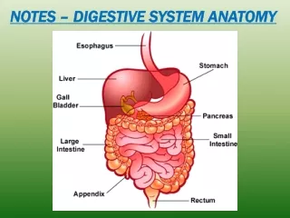
OESOPHAGUS Musculartube about 10 in. (25 cm) long Extendsfrom the pharynx to the stomach. Beginsin the neck where it is continuous with the laryngopharynx . Consistsof striated (voluntary) muscle in its upper 1/3, smooth (involuntary) muscle in its lower 1/3, and a mixture of striated and smooth muscle in between.
STOMACH Expandedpart of the digestive tract between the esophagus and small intestine. Specializedfor the accumulation of ingested food, chemically and mechanically prepares for digestion and passage into the duodenum. Actsas a food blender and reservoir; its chief function is enzymatic digestion.
The size, shape, and position of the stomach can vary markedly in persons of different body types (bodily habitus) May change even in the same individual as a result of Diaphragmaticmovements during respiration Stomach'scontents (empty vs. after a heavy meal) Positionof the person.
The stomach has four parts: Cardia: part surrounding the cardial orifice (opening), the superior opening or inlet of the stomach. Fundus:dilated superior part related to the left dome of the diaphragm and is limited inferiorly by the horizontal plane of the cardial orifice. Body:major part of the stomach between the fundus and pyloric part. Pyloric part: funnel-shaped outflow region of the stomach.
SMALL INTESTINE Primarysite for absorption of nutrients from ingested materials. Extendsfrom the pylorus to the ileocecal junction where the ileum joins the cecum (the first part of the large intestine).
Duodenumfirst part of the small intestine Shortest, widest and most fixed part. Jejunumbegins at the duodenojejunal flexure where the digestive tract resumes an intraperitoneal course. Ileumends at ileocecaljunction, union of the terminal ileum & cecum. Together, jejunum and ileum are 6-7 m long. Jejunum2/5 , Ileum 3/5 intraperitonealsection of the small intestine.
Most of the jejunum lies in the left upper quadrant (LUQ), whereas most of the ileum lies in the right lower quadrant (RLQ). The terminal ileum usually lies in the pelvis from which it ascends, ending in the medial aspect of the cecum.