Download
1 / 23
230 likes | 405 Views
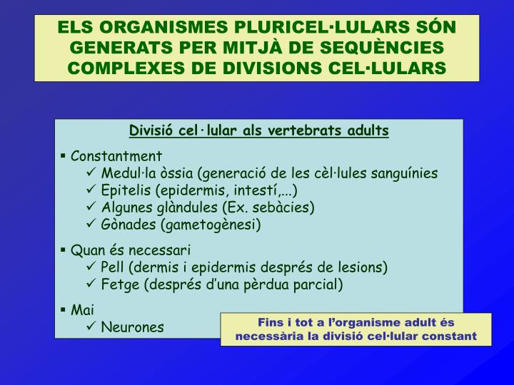
ELS ORGANISMES PLURICEL·LULARS SÓN GENERATS PER MITJÀ DE SEQUÈNCIES COMPLEXES DE DIVISIONS CEL·LULARS. Divisió cel·lular als vertebrats adults Constantment Medul·la òssia (generació de les cèl·lules sanguínies Epitelis (epidermis, intestí,...) Algunes glàndules (Ex. sebàcies)

E N D
ELS ORGANISMES PLURICEL·LULARS SÓN GENERATS PER MITJÀ DE SEQUÈNCIES COMPLEXES DE DIVISIONS CEL·LULARS • Divisió cel·lular als vertebrats adults • Constantment • Medul·la òssia (generació de les cèl·lules sanguínies • Epitelis (epidermis, intestí,...) • Algunes glàndules (Ex. sebàcies) • Gònades (gametogènesi) • Quan és necessari • Pell (dermis i epidermis després de lesions) • Fetge (després d’una pèrdua parcial) • Mai • Neurones Fins i tot a l’organisme adult és necessària la divisió cel·lular constant
FASES DEL CICLE CEL·LULAR Figure 13-1. The fate of a single parental chromosome throughout the eukaryotic cell cycle. Although chromosomes condense only during mitosis, they are shown in condensed form to emphasize the number of chromosomes at different cell-cycle stages. The nuclear envelope is not depicted. Following mitosis (M), daughter cells contain 2n chromosomes in diploid organisms and 1n chromosomes in haploid organisms including yeasts maintained in the haploid state. In proliferating cells, G1 is the period between “birth” of a cell following mitosis and the initiation of DNA synthesis, which marks the beginning of the S phase. At the end of the S phase, cells enter G2 containing twice the number of chromosomes as G1 cells (4n in diploid organisms). The end of G2 is marked by the onset of mitosis, during which numerous events leading to cell division occur. The G1, S, and G2 phases are collectively referred to as interphase, the period between one mitosis and the next. Most nonproliferating cells in vertebrates leave the cell cycle in G1, entering the G0 state.(Fuente: Lodish et al., 2000)
CONTROL DEL CICLE CEL·LULAR Figure 17-10. Two key components of the cell-cycle control system. A complex of cyclin with Cdk acts as a protein kinase to trigger downstream processes. Without cyclin, Cdk is inactive.(Fuente: Alberts et al., 1993) Figure 17-9. Checkpoints and inputs of regulatory information to the cell-cycle control system. Feedback from downstream processes and signals from the environment can prevent the control system from passing through certain specific checkpoints. The most prominent checkpoints are where the control system activates the triggers shown in yellow boxes. (Fuente: Alberts et al., 1993)
REGULACIÓ DEL CICLE CEL·LULAR Figure 13-2. Current model for regulation of the eukaryotic cell cycle. Passage through the cycle is controlled by G1, S-phase, and mitotic cyclin-dependent kinase complexes (CdkCs) highlighted in green. These are composed of a regulatory cyclin subunit and a catalytic cyclin-dependent kinase subunit. Protein complexes (orange) in the Cdc34 pathway and APC pathway polyubiquitinate specific substrates including the S-phase inhibitor, anaphase inhibitor, and mitotic cyclins, marking these substrates for degradation by proteasomes. These pathways thus drive the cycle in one direction because of the irreversibility of protein degradation. Proteolysis of anaphase inhibitors inactivates the protein complexes that connect sister chromatids at metaphase (not shown), thereby initiating anaphase.(Fuente: Lodish et al., 2000)
L’ESTUDI DEL CICLE CEL·LULAR ALS OVOCITS DE Xenopus VA PERMETRE DESCOBRIR EL FACTOR PROMOTOR DE LA MITOSI (MPF) Figure 17-15. Assaying for MPF by injection into a Xenopus oocyte. MPF can be detected because it drives the oocyte into M phase. The large nucleus (or "germinal vesicle") of the oocyte breaks down as the mitotic spindle forms. (Fuente: Alberts et al., 1993) • 12 primeres divisions en 7 hores (212 =4096) • Sense fases G1 i G2 • Sense a penes síntesi proteica (reserves) Peculiaritats de les primeres divisions als embrions de Xenopus
LA EXPRESSIÓ DEL MPF ÉS CÍCLICA, IGUAL QUE LA D’UNES PROTEÏNES DENOMINADES CICLINES Figure 17-19. Rise and fall in levels of MPF and cyclin during the early embryonic cell cycle. The cyclin measurements have been made chiefly in the eggs of marine invertebrates, where cyclin accounts for 5% of the protein synthesized during a brief pulse with radioactive amino acids. The gels below the graph show the amounts of labeled cyclin of two varieties, A and B, at different stages of the cycle of a clam egg. The bottom line in the gel shows the synthesis of a house-keeping enzyme that serves as a standard of comparison. (Adapted from T. Hunt, F.C. Luca, and J.V. Ruderman, J. Cell Biol. 116:707-724, 1992.) (Fuente: Alberts et al., 1993)
L’EXTRACTE DE CITOPLASMA D’UN OVOCIT ACTIVAT ÉS CAPAÇ D’INDUIR LA MITOSI DINS D’UN TUB D’ASSAIG Aquest tipus d’experiments in vitro han permès demostrar que la síntesi i la degradació de les ciclines està correlacionada amb l’activitat del MPF i els esdeveniments mitòtics (veure diapositiva següent) Figure 17-20. Cycling in a cell-free system. A large batch of activated frog eggs are broken open by gentle centrifugation, which also separates their cytoplasm from other components. The undiluted cytoplasm is collected, and sperm nuclei are added to it, together with ATP. The sperm nuclei decondense and then go through repeated cycles of mitosis and DNA replication, indicating that the cell-cycle control system is operating in this cell-free cytoplasmic extract. (Fuente: Alberts et al., 1993)
RELACIÓ ENTRE LA CONCENTRACIÓ DE CICLINA, L’ACTIVITAT DEL MPF I LA MITOSI EN EXPERIMENTS IN VITRO Figure 13-7. Experimental demonstration that the synthesis and degradation of cyclin B are required for the cycling of MPF activity and mitotic events in Xenopus egg extracts. In all cases, MPF activity and cyclin B concentration were determined at various times after addition of sperm chromatin to an extract treated as indicated. Microscopic observations determined the occurrence of early mitotic events (blue shading), including chromosome condensation and nuclear envelope breakdown, and of late events (orange shading), including chromosome decondensation and nuclear envelope reformation. See text for discussion. [See A. W. Murray et al., 1989, Nature339:275; adapted from A. Murray and T. Hunt, 1993,The Cell Cycle: An Introduction, W. H. Freeman and Company.](Fuente: Lodish et al., 2000)
ELS LLEVATS SÓN MODELS MOLT ÚTILS PER A L’ESTUDI GENÈTIC DEL CONTROL DEL CICLE CEL·LULAR Figure 17-24. A comparison of the cell cycles of fission yeast and budding yeast. The fission yeast shown in the upper panel has a typical eucaryotic cell cycle with G1, S, G2, and M phases. Unlike that of higher eucaryotic cells, however, the nuclear envelope of the yeast cells does not break down: the microtubules of the mitotic spindle (green) form inside the nucleus and are attached to spindle pole bodies (dark green) at its periphery. The cell divides by forming a partition (known as the cell plate) and splitting in two. The budding yeast has normal G1 and S phases. However, a microtubule-based spindle begins to form very early in the cycle, during S phase; thus there does not appear to be a normal G2 phase. In contrast with fission yeasts, the cell divides by budding. As in fission yeasts, but in contrast with higher eucaryotic cells, the nuclear envelope remains intact during mitosis. The condensed mitotic chromosomes (red) are readily visible in fission yeasts, but are less easily seen in budding yeasts (Fuente: Alberts et al., 1993)
L’ESTUDI DE DIFERENTS LLEVATS MUTANTS HA PERMÉS CARACTERITZAR MOLECULARMENT ELS PUNTS DE CONTROL DEL CICLE CEL·LULAR Figure 17-27. Genesis of MPF activity. Cdc2 becomes associated with cyclin as the level of cyclin gradually increases; this enables Cdc2 to be phosphorylated by an activating kinase on an "activating" site as well by Wee1 kinase on Cdc2's catalytic site. The latter phosphorylation inhibits Cdc2 activity until this phosphate group is removed by the Cdc25 phosphatase. Active MPF is thought to stimulate its own activation by activating Cdc25 and inhibiting Wee1, either directly or indirectly. (Fuente: Alberts et al., 1993) Figure 17-30. The Cdc2 cycle in yeast. Cdc2 is permanently present, but its state of association with cyclins changes, defining the division-cycle phase of the cell. (Fuente: Alberts et al., 1993)
ALS MAMÍFERS S’HAN TROBAT VARIES FORMES DE CICLINES I DE CDKs Figure 13-29. Activity of mammalian Cdkcyclin complexes through the course of the cell cycle in G0 cells induced to divide by treatment with growth factors. The width of the colored bands is approximately proportional to the protein kinase activity of the indicated complexes. Cyclin D refers to all three D-type cyclins. (Fuente: Lodish et al., 2000)
EL CICLE CEL·LULAR ÉS INDUÏT ALS MAMÍFERS PER FACTORS DE CREIXEMENT
ELS FACTORS DE CREIXEMENT ACTIVEN LA TRANSCRIPCIÓ DE GENS D’EXPRESSIÓ IMMEDIATA Figure 17-45. The response of Myc to a growth factor. Myc is the product of the early-response gene myc. The graph shows the changes in the concentration of Myc protein following a sudden increase in growth factor concentration to a new steady value, which causes the cell to exit G0 and to proliferate. The changes in Myc concentration reflect changes in myc gene transcription, stimulated by exposure of the cell to the growth factor. Myc protein itself inhibits myc transcription, and this negative feedback is thought to explain why the level of Myc declines from its initial peak to a lower steady value. (Fuente: Alberts et al., 1993) Un altre gen d’expressió immediata és c-fos, el qual s’utilitza com a marcador d’activitat cel·lular a cèl·lules en G0, com neurones
ELS GENS D’EXPRESSIÓ IMMEDIATA SÓN PROTOONCOGENS. LES SEUES MUTACIONS PODEN ORIGINAR CÀNCER Figure 17-43. Tumor-suppressor genes versus proto-oncogenes. The product of a tumor-suppressor gene inhibits assembly and activation of the cell-cycle control system; the product of a proto-oncogene does the opposite. Unrestrained proliferation can result from mutations that either inactivate both copies of the tumor-suppressor gene or strongly overactivate one copy of the proto-oncogene. (Fuente: Alberts et al., 1993) Però també hi ha gens que actuen com a gens supressors de tumors
LA PROTEÏNA RETINOBLASTOMA EVITA L’ACCIÓ DE FACTORS DE TRANSCRIPCIÓ QUE ACTIVEN LA SÍNTESI DE PROTEÏNES NECESSÀRIES PER LA FASE S (E2F) (CICLINA S I ALTRES) Figure 17-46. Action of the retinoblastoma (Rb) protein. Dephosphorylated Rb binds to, and holds inactive, gene regulatory proteins that stimulate transcription of target genes (such as myc) required for cell proliferation. Phosphorylated Rb detaches, releasing the stimulatory proteins that activate proliferation. (Fuente: Alberts et al., 1993)
ALGUNES CARACTERÍSTIQUES DE LA FASE M • Condensació de la cromatina en cromosomes – fosforilació de condensines per M-Cdk • Vesiculació de la membrana nuclear - fosforilació de la làmina nuclear per M-Cdk • Augment de la inestabilitat dels microtúbuls que permet la organització del fus mitòtic - Fosforilació de Proteïnes Associades a Microtúbuls • Els orgànuls amb membrana (RE, Golgi) també es trenquen en vesícules. Es deté el trànsit vesicular • Duplicació del centrosoma. Sembla que el procés és activat per la ciclina E-Cdk2 en mamífers Figure 18-4. Centriole replication. The centriole pair is associated with the centrosome matrix (green). At a certain point in G1 phase the two centrioles separate by a few micrometers. During S phase a daughter centriole begins to grow near the base of each old centriole and at a right angle to it. The elongation of the daughter centriole is usually completed by G2 phase. The two centriole pairs remain close together in a single centrosomal complex until the beginning of M phase, when the centrosome splits in two and the two halves begin to separate. (Fuente: Alberts et al., 1993)
L’APARELL MITÒTIC ÉS UNA MÀQUINA DE MICROTÚBULS PER SEPARAR CROMOSOMES Figure 19-37. Mitotic apparatus in S. cerevisiae. In yeast, the nucleus remains intact during mitosis; thus the chromosomes are isolated from direct interaction with the cytosol. (a) Spindle pole bodies, which are attached to the nuclear membrane, organize the spindle microtubules (MTs). Each of the 16 chromosomes is attached to two kinetochore microtubules. (Fuente: Lodish et al., 2000) Figure 19-36. Diagram showing the three sets of microtubules (MTs) in the mitotic apparatus. Centered around the poles are astral microtubules, kinetochore microtubules, which are connected to chromosomes (blue), and polar microtubules. The (+) ends of these microtubules all point away from the centrosome at each pole. (Fuente: Lodish et al., 2000)
ESTRUCTURA DEL CINETOCOR I EL CENTRÒMER Figure 19-39. Centromeric attachment of microtubules. (a) Schematic diagram of attachment of kinetochore microtubules to the sister chromatids of a metaphase chromosome. In animals and lower plants, the kinetochore is a three-layer, platelike structure lying within the centromere of each chromatid (inset). The (+) ends of microtubules insert into the outer layer of each kinetochore, and the microtubules extend toward one of the two poles of the cell. At anaphase, the sister chromatids separate, and the chromosomes are pulled to opposite poles of the cell by the kinetochore microtubules. (b) Association of a yeast centromere with components of the kinetochore. The centromeric (CEN) DNA is divided into three contiguous segments (CDE I – III). Two groups of centromere-binding factors, CBF2 and CBF3, are proteins associated with CDE II and CDE III, respectively. The CBFs mediate the attachment of a single microtubule to the centromere. Other microtubule-binding proteins form the rest of the kinetochore. [Part (a) adapted from A. F. Pluta et al., 1995, Science270:1591.] (Fuente: Lodish et al., 2000)
PROTEÏNES MOTORES I ORGANITZACIÓ DEL FUS MITÒTIC Figure 19-43. Spindle pole formation. Model of the interactions between spindle microtubules (MTs) and various proteins at the pole. [Adapted from T. Gaglio et al, 1995, J. Cell Biol.135:399] (Fuente: Lodish et al., 2000) Figure 19-42. Model for participation of microtubule motor proteins in centrosome movements during mitosis. (a) During late prophase centrosomes are aligned by (−) end – directed motors (dark green), which pull on polar microtubules and thus align them. (b) A family of (+) end – directed KRPs (pink), probably BimC kinesins, associated with the polar microtubules is involved in separation of the poles — beginning after centrosome alignment, continuing through the formation of the spindle poles, and ending, as we will see later, with spindle pole separation at anaphase. In addition, a (−) end – directed force exerted by cytosolic dynein (light green) located at the cortex may pull asters toward the poles in combination with the (+) end – directed motors in the spindle. (Fuente: Lodish et al., 2000)
ELS CINETOCORS S’UNEIXEN ALS EXTREMS + DELS MICROTÚBULS DEL FUS MITÒTIC Figure 19-44. Dynamic instability and the capture of chromosomes. (a) During mitotic prophase, some spindle microtubules are growing at their distal (+) end, while others are shrinking rapidly. (b) In late prophase, the ends of some microtubules interact with kinetochores (dark green), causing those microtubules to be stabilized. (c) In addition, some microtubules just miss the kinetochore, but the kinetochore binds to the side of the microtubule and then slides to the (+) end. (Fuente: Lodish et al., 2000)
ELS CROMOSOMES ES MANTENEN EN LA PLACA METAFÀSICA GRÀCIES A FORCES BIPOLARS EQUILIBRADES Figure 19-45. Proposed alternative mechanisms for chromosome congression. A coupling of microtubule dynamics and microtubule motors may keep a chromosome positioned at the equator. (a) The flow of tubulin subunits through kinetochore microtubules can be used to pull or push the chromosome relative to the pole, especially if rapid polymerization or depolymerization occurs at the (+) end of a microtubule. (b) A (−) end–directed motor (dark green) at the kinetochore or a (+) end–directed motor (pink) at the spindle pole can pull a chromosome toward the pole, while CENP-E tethers the kinetochore to a shrinking microtubule. (c) Nonkinetochore microtubules can exert a pushing force on the chromatid arms of a chromosome by polymerization at their (+) ends. (Fuente: Lodish et al., 2000)
DURANT L’ANAFASE A ELS MICROTÚBULS CINETOCÒRICS ES DESMUNTEN Tallar amb làser Els microtúbuls quinetocòrics es despolimeritzen als extrems positius i els quinetocors es mouen cap als pols Figure 19-46. Experimental demonstration that during anaphase A chromosomes move poleward along stationary kinetochore microtubules, which coordinately disassemble from their kinetochore ends. Fibroblasts are injected with fluorescent tubulin and then allowed to enter metaphase, so that all the microtubules are fluorescent. Only the kinetochore microtubules are shown. In early anaphase, a band of microtubules (yellow box) is subjected to a laser light, which bleaches the fluorescence but leaves the microtubules continuous and functional across the bleached region. The bleached segment of each microtubule thus provides a marker for the position of that part of the microtubule. During anaphase the distance of the bleached zone from the poles (measured in the diagrams by the black double-headed arrows) does not change, indicating that no depolymerization of the microtubules occurs at the poles. Rather, the kinetochore microtubules disassemble just behind the kinetochore, and the kinetochores move poleward along the microtubules. [Adapted from G. J. Gorbsky et al., 1987, J. Cell Biol.104:9; and G. J. Gorbsky et al., 1988, J. Cell Biol.106:1185.] (Fuente: Lodish et al., 2000)
DURANT L’ANAFASE B ELS MICROTÚBULS POLARS RELLISQUEN UNS SOBRE ALTRES (PER MIG DE PROTEÏNES MOTORES) ALLARGANT EL FUS MITÒTIC Figure 19-47. Model of spindle elongation and movement during anaphase B. Tubulin (light purple) adds to the (+) ends of all polar microtubules, lengthening these fibers. Simultaneously, (+) end – directed KRPs (pink) bind to the polar microtubules in the overlap region. Each KRP, bound to a microtubule in one half-spindle, “walks” along a microtubule in the other half-spindle, toward its (+) end, utilizing the energy from ATP hydrolysis. In cells that assemble an aster, a (−) end – directed motor protein (light green) located in the cortex of the plasma membrane pulls on the astral microtubules, which also moves the poles farther apart. [Adapted from H. Masuda and W. Z. Cande, 1987, Cell49:193.] (Fuente: Lodish et al., 2000) Els microtúbuls astrals contribueixen estirant dels centrosomes cap als pols