Download
1 / 64
640 likes | 740 Views
This chapter outlines the key aspects of the animal body, including epithelial tissue characteristics, types of skeletons, bone structure, muscle actions, and the sliding filament mechanism. It explores the organization of the body, from cells and tissues to organs and organ systems. The unique properties of epithelial tissue, such as regeneration and barrier functions, are detailed, along with the different types of epithelial tissues and glands. Additionally, it covers connective tissues like loose and dense connective tissues, special connective tissues like cartilage and bone, and muscle tissue types. The formation and structure of bone, as well as the classification of blood as a connective tissue, are also discussed in this comprehensive overview.

E N D
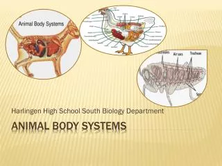
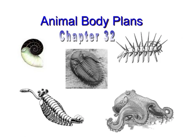
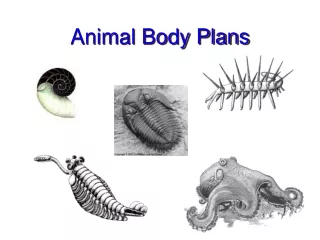
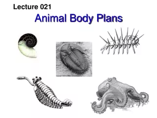
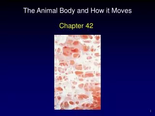
The Animal Body and How it Moves Chapter 42
Outline • Characteristics of Epithelial Tissue • Tissue Types • Types of Skeletons • The Structure of Bone • Types of Joints • Actions of Skeletal Muscles • Sliding Filament Mechanism of Contraction • Control of Muscle Contraction • Types of Muscle Fibers • Modes of Animal Locomotion
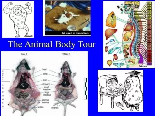
Organization of the Body • Bodies of all vertebrates are basically a tube within a tube. • all vertebrate bodies supported by internal skeleton • Four levels of organization: • cells • tissues • organs • organ systems
Organization of the Body • Tissues • Groups of cells similar in structure and function are organized into tissues. • Early in development, embryo cells differentiate into three germ layers. • endoderm • mesoderm • ectoderm
Tissues • Adult vertebrates have four primary tissues: • epithelial • connective • muscle • nerve
Organization of the Body • Organs and organ systems • Organs are body structures composed of several different tissues that form a structural and functional unit. • An organ system is a group of organs that operate to perform the major activities of the body.
Characteristics of Epithelial Tissue • Epithelium covers every major surface of the vertebrate body. • derived from all three germ layers • can provide a barrier that can impede the passage of some substances while facilitating the passage of others • remarkable regenerative powers
Characteristics of Epithelial Tissue • Types of epithelial tissues • simple - one layer thick • squamous - lining of lungs • cuboidal - lining of kidney tubules • columnar - lining of stomach • stratified - several cell layers thick and named according to features of their uppermost layers
Characteristics of Epithelial Tissue • Glands of vertebrates are derived from invaginated epithelium. • exocrine glands - connection between the gland and the epithelial membrane is maintained as a duct • endocrine glands - ductless glands - connections with the epithelium, from which they are derived, are lost during development • secrete hormones
Connective Tissue Proper • Connective tissues are divided into: • connective tissue • divided into loose and dense connective tissues • special connective tissues • include cartilage, bone, and blood • extracellular material generically known as matrix
Connective Tissue Proper • Loose connective tissue • cells scattered within amorphous mass of proteins that form a ground substance • strengthened by collagen, elastin and reticulin - secreted by fibroblasts • adipose cells found in loose connective tissue
Connective Tissue Proper • Dense connective tissue • regular • collagen fibers lined up in parallel • tendons and ligaments • irregular • collagen fibers have many orientations • organ coverings - capsules • muscle coverings - epimysium • nerve coverings - perineurium • bone covering - periosteum
Special Connective Tissues • Cartilage • specialized connective tissue in which fibers are laid down along the lines of stress in long, parallel arrays • firm and flexible • chondrocytes - cartilage cells that live within spaces (lacunae) within cartilage matrix
Special Connective Tissues • Bone • Many bones are first modeled in cartilage. The cartilage matrix calcifies at particular locations, thus chondrocytes are no longer able to obtain oxygen and nutrients through diffusion.
The Structure of Bone • New bone is formed by osteoblasts that secrete collagen organic matrix in which calcium phosphate is later deposited. • cells then encased in spaces called lacunae in the calcified matrix • Bone is constructed in thin, concentric layers or lamellae, laid down around Haversian canals that run parallel to the length of the bone. • contain nerve fibers and blood vessels
The Structure of Bone • Bone formation • flat bones - Osteoblasts located in a web of dense connective tissue produce bone within that tissue. • long bones - bone first “modeled” in cartilage • ends and interior composed of spongy bone
Special Connective Tissues • Blood • classified as connective tissue because it contains plasma and platelets • erythrocytes - contain hemoglobin • leukocytes - have nuclei and mitochondria, but lack hemoglobin • neutrophils, eosinophils, and basophils • lymphocytes and monocytes
Muscle Tissue • Muscle cells are the motors of the vertebrate body. • three types: smooth - skeletal - cardiac • Skeletal and cardiac muscles are striated because their cells have transverse stripes when viewed in longitudinal section. • Contraction of skeletal muscle is under voluntary control, whereas contraction in cardiac and smooth muscle is generally involuntary.
Muscle Tissue • Smooth muscle - found in organs of internal environment (viscera) • Skeletal muscle - usually attached to tendons or bones, so when muscles contract causes bones to move at joints • made up of long muscle fibers that contract by myofibrils • made up of highly ordered arrays of actin and myosin filaments
Muscle Tissue • Cardiac muscles • composed of smaller, interconnected cells, each with a single nucleus • interconnections appear as dark lines called intercalated disks • enable cardiac muscles to form single functioning unit - myocardium
Nerve Tissue • Cells include neurons and neuroglia (supporting cells). • Neurons are specialized to produce and conduct electrochemical impulses.
Nerve Tissue • Neuroglia do not conduct electrical impulses but instead support and insulate neurons and eliminate foreign materials in and around neurons. • myelin sheath - insulating covering of neuroglia cells wrapped around axons • nodes of Ranvier separate adjacent neuroglia cells
Nerve Tissue • Nervous system is divided in the central nervous system (CNS) which includes the brain and spinal cord, and the peripheral nervous system (PNS) which includes nerves and ganglia. • Nerves consist of axons in the PNS bundled together. • Ganglia are collections of neuron cell bodies.
Types of Skeletons • Hydrostatic skeletons - fluid-filled cavity encircled by muscle fibers • As the muscles contract, fluid in the cavity moves and changes cavity shape. • Exoskeletons - surround the body as a rigid, hard case • must be periodically shed • limits body size as exoskeleton has to grow increasingly thicker and heavier
Types of Skeletons • Endoskeletons - rigid internal skeleton to which muscles are attached • composed of cartilage or bone • vertebrate skeleton • axial skeleton - forms axis of body and supports organs of the head, neck, and chest • appendicular skeleton - includes bones of the limbs, pectoral and pelvic girdles
Actions of Skeletal Muscles • Skeletal muscles produce movement of the skeleton when they contract. • attachment to bones made by tendons • origin remains stationary during contraction • insertion attached to bone that moves during contraction
Actions of Skeletal Muscles • Synergists - muscles that cause same action at a joint • Antagonists - muscles that produce opposing actions • Isotonic contraction - muscle and all fibers shorten in length thus force of contraction remains relatively constant • Isometric contraction - tension is absorbed by tendons and other elastic tissue, and muscle does not change in length
Sliding Filament Mechanism of Contraction • Each skeletal muscle contains numerous muscle fibers. • Each muscle fiber encloses 4-20 myofibrils. • Each myofibril composed of thick and thin myofilaments. • Thick myofilaments produce A bands. • Thin myofilaments produce I bands. • Each I band divided in half by disc of protein (Z band).
Sliding Filament Mechanism of Contraction • Sarcomere - structure of myofibril from Z line to Z line • smallest subunit of muscle contraction • A muscle contracts and shortens because its myofibrils contract and shorten. • Myofilaments do not shorten, but slide deeper into the A band.
Sliding Filament Mechanism of Contraction • Electron micrographs reveal cross-bridges that extend from the thick to thin filaments. • Each thick filament composed of many myosin proteins packed together, and every myosin molecule has a “head” region. • Each thin filament consists primarily of many globular actin proteins twisted in a double helix.
Sliding Filament Mechanism of Contraction • Before the myosin heads bind to the actin of the thin filaments, they act as ATPase, splitting ATP into ADP and Pi. • activates heads • Once a myosin head binds to actin, it undergoes a shape change, pulling the thin filament toward the center of the sarcomere. • allows head to detach from actin and continue cross-bridge cycle
Control of Muscle Contraction • Role of Ca++ in contraction • When a muscle is relaxed the myosin head cannot bind to actin because the attachment sites are physically blocked by tropomyosin. • In order to contract a muscle, troponin must move tropomyosin away from the binding site. • complex regulated by calcium ion concentration
Control of Muscle Contraction • When Ca++ concentration of the muscle cell cytoplasm is low, tropomyosin inhibits cross-bridge formation and the muscle is relaxed. • When Ca++ concentration is raised, Ca++ binds to troponin. • When a muscle fiber is stimulated to contract, an electrical impulse travels into the muscle fiber down transverse tubules. • triggers release of Ca++ from sarcoplasmic reticulum
Control of Muscle Contraction • Nerves stimulate contraction • Somatic motor neurons stimulate skeletal muscles. • Axon extends from neuron cell body and branches to make synapses with a number of muscle fibers.
Control of Muscle Contraction • Somatic motor neuron stimulates contraction: • releasing acetylcholine neurotransmitter (ACh). • impulses spread along membrane and carried into the muscle fibers through the T tubules • T tubules conduct impulse toward the sarcoplasmic reticulum, which releases Ca++ • Excitation-contraction coupling
Control of Muscle Contraction • Motor units and recruitment • set of muscle fibers innervated by all axonal branches is defined as a motor unit • division of muscle into motor units allows muscle’s strength of contraction to be finely graded • most muscles contain motor units in a variety of sizes • recruitment - nervous system’s use of increased numbers and sizes of motor units to produce a stronger contraction
Types of Muscle Fibers • Muscle fiber twitches • muscle stimulated with a single electric shock • A second electrical shock delivered immediately after the first will produce a second twitch that may partially piggyback on the first (summation). • At a particular frequency of stimulation, there is no visible relaxation between successive twitches (tetanus).