Download
1 / 72
720 likes | 742 Views
Explore the intricate levels of structure within the human body, from cells to tissues and organ systems. Learn about epithelial and connective tissue types, their functions, and special connective tissues like cartilage and bone. Discover how muscle and nerve tissues support movement and transmit impulses. This guide offers insights into the diverse functions of each tissue type and their vital roles in maintaining the body's health.

E N D
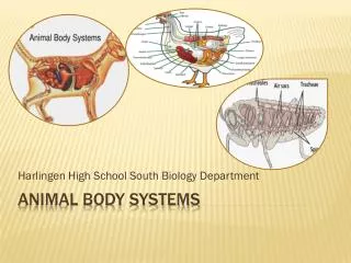
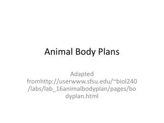
Levels of structure • Cells • Tissues: Cells combined in distinct ways into layers. Specialization (division of labor) • Organs: Made of more than one kind of tissue. Perform common function. • Organ systems: Several organs combine to perform major body function.
Levels of structure • Example, circulatory system
Levels of structure • 11 organ systems
Tissue types (4) • 1) Epithelial tissue • Covers outer and inner ____________ of body • Cells tightly joined together (no blood vessels in intercellular spaces: materials must diffuse in and out) • Two major types: covering and lining, and glandular
Tissue types • 1) Epithelial tissue types (covering and lining) • Simple epithelial (1 cell layer). Further subdivided based on cell shape • Squamous (flattened) • Cuboidal (cube shaped) • Columnar (rectangular)
Tissue types • 1) Epithelial tissue types (covering and lining) • Stratified epithelial (multiple cell layers). Further subdivided by cell shape • Ex., squamous (flattened) • Pseudostratified epithelial (one cell layer but looks like 2 because __________ not all in same position) • Ex., pseudostratified columnar (rectangular)
Tissue types • 1) Epithelial tissue • Glandular (secretes materials) epithelial tissue • Exocrine glands: connected to epithelium surface by duct (Ex., sweat glands, salivary glands) • Endocrine glands: not connected to epithelium surface (Ex., adrenal gland of kidney). Secretions called hormones, enter blood and stay within body
Tissue types • Epithelial tissue functions: • Protection: invasion, dehydration, physical injury, etc. • Secretion of chemicals: ex., digestive tract • Note: usually with great regenerative ability • Ex., stomach lining replaced every ______ days
Tissue types • 2) Connective tissue • Used to bind and support all other tissues • Other special functions too • Composed of cells and matrix (extracellular material) • Two types: • Connective tissue proper • Special connective tissues
Tissue types • Connective tissue proper • 1) Loose connective tissue • Matrix often contains: collagen (strong protein fibers), elastin (elastic protein fibers)
Tissue types • Connective tissue proper • 1) Loose connective tissue • Cells include: ______________ (cells that secrete matrix), mast cells (make histamine: blood vessel dilator), macrophages (phagocytic cells), adipose cells (contain a fat droplet) Adipose Loose connective
Tissue types • Connective tissue proper • 2) Dense connective tissue • Collagen fibers tightly packed (very strong) • Ex, tendons and ligaments
Tissue types • Special connective tissues: cartilage • Matrix: collagen fibers in parallel arrays and glycoproteins
Tissue types • Special connective tissues: cartilage • Chondrocytes: living cartilage cells. Live in chambers (lacunae). Note lack of blood vessels: diffusion supplies cells with materials (slow process!)
Tissue types • Special connective tissues: bone • Matrix hardened with calcium phosphate salts • No __________ occurs through bone matrix. Blood vessels present in central canals
Tissue types • Special connective tissues: bone • Cells called osteocytes, located in chambers (lacunae) • Osteocytes connect with cellular processes through canalaliculi
Tissue types • Special connective tissues: blood • Blood is connective tissue, with fluid matrix (plasma) • Erythrocytes (red blood cells): carry oxygen and carbon dioxide • Leukocytes (white blood cells): ___________ cells • Thrombocytes (platelets): fragments of bone marrow cells
Tissue types • Connective tissue functions: • Connect parts (tendons connect muscle to bone) • Support body (skeleton) • Protection (cranium around brain) • Circulate materials (blood).
Tissue types • 3) Muscle tissue • Most abundant _________ in humans • Basic structure: bundles of myofibrils, composed of protein filaments (actin and myosin).
Tissue types • 3) Muscle tissue types: • Skeletal (striated) muscle: multinucleated, produce voluntary movements
Tissue types • 3) Muscle tissue types: • Cardiac (also striated): single nucleus, have intercalated discs (heart muscle)
Tissue types • 3) Muscle tissue types: • Smooth muscle: no striations. Usually not under voluntary control
Tissue types • Functions: • Movement (of body, fluids within body, etc.)
Tissue types • 4) Nerve tissue • Transmits electrochemical impulses • Neuron (name of cell type) • Dendrite (process: brings impulse to cell body) • Axon (process: carries impulse from cell body)
Tissue types • 4) Nerve tissue • Neuroglia: support cells • Often associate with axons, form sheath (_________ sheath) • Note Nodes of Ranvier (gaps in sheath).
Tissue types • 2 nerve networks: • Peripheral Nervous System (PNS): perceives environment and communicates to body • Central Nervous System (CNS): processes information and coordinates activities
Tissue types • 4) Nerve tissue: Types of neurons • Sensory (receive information: send to CNS) • Motor (stimulate muscles/glands) • Association (integrate information and connect to other neurons)
Tissue types • 4) Nerve tissue • Nerves: bundles of ___________ • Ganglion (plural: ganglia): collection of neuron cell bodies
Tissue types • 4) Nerve tissue • Functions: Send electrical signals to convey information, make things happen in body, etc.
Integumentary System • Human skin: largest organ of body (2 square yards, 16% of body weight) • Distribution: “Rule of _____s” Note: diagram %s are for only one side of body
Integumentary System • Many functions in animals: • Protection. Forms boundary of individual, barrier to external dangers and valuable internal materials • Sensing. Can gather information about environment: temperature, pressure, light, damage to integument • Communication. Since visible to others, can send signals with skin color or structures (hairs, scales, feathers) • Regulate body temperature. Can be used to gain/lose energy. Integument structures (hairs, scales, feathers) can provide ____________
Integumentary System • Many functions in animals: • Excretion: Sweat contains water, urea (nitrogenous waste material), salts. These eliminated during sweating
Integumentary System • Skin layers • Epidermis • Dermis
Integumentary System • Human skin layers: Epidermis • Upper boundary: Stratum corneum. Dead cells that provide protective layer. Shed as skin scales or flakes
Integumentary System • Human skin layers: Epidermis • You will shed about 40 lbs. of skin scales during your lifetime • Good news for dust mites (Phylum Arthropoda, Class Arachnida, Order Acari)!
Integumentary System • Human skin layers: Epidermis • Lower boundary: Stratum germinativum (basal cells). Living cells doing rapid _________. Daughter cells fill with keratin (tough water resistant protein: also found in hair, fingernails, hooves, claws), flatten and die
Integumentary System • Human skin layers: Epidermis • Contains melanocytes: cells that produce melanin and transfer it to other skin cells. Darken skin color in response to UV exposure (tanning)
Integumentary System • Human skin layers: Epidermis • UV exposure increases skin cancer chances • Melanoma: ____________ that divides out of control • Recall ozone thinning problem
Integumentary System • Human skin layers: Dermis • Contains connective tissue, blood vessels, nerve endings.
Integumentary System • Human skin also contains: hairs • Follicle: invagination of skin surface containing hair • Shaft: body of hair (mostly protein: ____________) • Bulb: base from which hair grows by cell division (each cell divides every 1-3 days!) • Papilla: contains blood supply
Integumentary System • Human skin also contains: hairs • Arrector pili muscle: smooth muscle. Can change angle of hair
Integumentary System • Human skin also contains: exocrine glands • Sweat glands: empty onto skin surface at sweat pore.
Integumentary System • Human skin also contains: exocrine glands • Sebaceous glands: associated with hair follicle, secrete oil to lubricate/protect skin and hair
Integumentary System • Human skin also contains: sense organs • Free nerve endings: sense _____________ • Hair follicle receptors: can sense movement of hair
Integumentary System • Human skin also contains: sense organs • Merkel cell, Meissner’s corpuscles, Pacinian corpuscles. Sense pressure/stretch of skin
Integumentary System • Human skin also contains: sense organs • Temperature: End-bulb of Krause (cold), Organ of Ruffini (hot)
Integumentary System • Below the skin: Subcutaneous __________ • Contains adipose tissue • Stores energy, cushioning (soles of feet, palms of hands), provides insulation
Integumentary System • Nails: Special keratinized layer of cells produced by fold of skin (nail root) Fingernail magnified 1000 X