Cardiac
Cardiac. Community College of Philadelphia Nursing 132 Spring 2007. Anatomy and Physiology Review. Structures Chambers Valves Arteries Physiology Direction of flow Preload Afterload. Coronary Circulation. Structure Epicardium Myocardium Endocardium Chambers Right and Left Atria

Cardiac
E N D
Presentation Transcript
Cardiac Community College of Philadelphia Nursing 132 Spring 2007
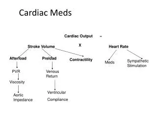
Anatomy and Physiology Review • Structures • Chambers • Valves • Arteries • Physiology • Direction of flow • Preload • Afterload
Structure • Epicardium • Myocardium • Endocardium • Chambers • Right and Left Atria • Right and left Ventricles • Valves • Atrioventricular • Tricuspid - Separates RA from RV • Mitral - Separates LA from the LV • Semilunar • Pulmonic – Separates RV from the Pulmonary Arteries • Aortic – Separates LV from the Aorta
Physiology • Blood flow through the heart • http://www-medlib.med.utah.edu/kw/pharm/hyper_heart1.html • Cardiac conduction Automaticity
Electrophysiology • Nodes • Pathway • Automaticity
Basic EKG Interpretation • What is an EKG and what does it measure/record? • PQRST • Measuring boxes • Horizontal measure time: small box 0.04 seconds Large box 0.20 seconds • Vertical voltage: small box 1mm or 0.1 mV large box 5mm or 0.5 mV
P wave • Atrial depolarization • Small, smooth, rounded • No taller than 2.5 mm • No wider than 0.11 sec
Q wave • First downward deflection • Should be less than 0.03 seconds in duration and less than 25% of the R wave • Indicates myocardial infarction
QRS Complex • Represents ventricular depolarization
T wave • Ventricular repolarization • Usually rounded • Should be same direction as QRS • Inverted T waves can be a sign of ischemia • Peaked T waves can be a sign of hyperkalemia
U wave • Small rounded wave not always present, thought to be part of ventricular repolarization
Angina (Stable Chronic) • Most common sign of ischemic heart disease • Myocardial oxygen demand is increased by exercise, smoking, eating heavy foods, weather extremes, emotional distress, etc… • Atherosclerosis causes progressive fixed narrowing of the arterial lumen • Relieved by rest or pharmacological interventions • Goal is to prolong survival, reduce disease progression
Acute Coronary Syndrome (ACS) • Unstable Angina (UA) • Non-ST Elevated Myocardial Infarction (NSTEMI) • ST Elevation Myocardial Infarction (STEMI) Penumbra
Unstable Angina (UA) / Non-ST Elevated Myocardial Infarction (NSTEMI) • Imbalance between myocardial oxygen supply and demand • UA occurs at rest without exertion • Reduced myocardial perfusion • Release of biochemical markers vs. no release of biochemical markers
Evaluation and management • Stratification High risk Intermediate Low risk
Immediate Management • History, PE, 12 – Lead EKG, initial cardiac markers • Assign to 1 of 4 categories • Definite or Possible • Cont. EKG monitoring • Cardiac markers • In facility observation • Repeat EKG and cardiac markers 6-12 hours
EKG and cardiac markers normal follow up stress test as outpatient acceptable • Definite ACS admit to hospital – Chest pain unit if available • Hospital Care • Nursing Management • Minimize or eliminate ischemia • Administer meds • Educate • Discharge teaching
ST Elevation Myocardial Infarction (STEMI) • MI occurs as a result of thrombotic occlusion of one or more of the coronary arteries • ******* • CP is most common symptom, severe, doesn’t go away • Diagnosis • EKG • Cardiac markers • PE, History
Ventricular Remodeling – Changes in the size, shape, and thickness of the left ventricle involving both the infarcted and non-infarcted segments of the ventricle. • Penumbra • 1.68 million unique discharges for ACS in 2001, 30% estimate to have STEMI
Management • Pt contact with healthcare system • Initiation of fibrinolytic therapy – < 30 minutes • Balloon inflation for PCI - < 90 minutes
Choice of treatment decided by EM physician and resources of institution and surrounding institutions • Contraindications to fibrinolytics
12 lead EKG completed and shown to experienced emergency physician WITHIN 10 minutes • Cardiac Markers • Troponin • CK-MB • Myoglobin • Lab results should not delay treatment
What should be done? • EKG • Lab work • Portable CXR • Oxygen • Nitroglycerin: 3 sublingual 0.04mg, IV if needed **** Phosphodiesterase inhibitor **** • Morphine Sulfate: 2 – 4mg IV • ASA chewed • Beta blocker if not contraindicated • ACE orally within 24 hours of STEMI for anterior infarct, pulm. congestion, LV EF < 40%
Patient Education Before Discharge • Lifestyle changes • Recognizing cardiac symptoms: calling 911 if symptoms not improving • Family education about AED, CPR • Lipid Management • Weight Management • Smoking cessation
Cont. • Antiplatelet Therapy: ASA, Plavix • ACE inhibitor if without contraindications • Beta blockers: except low risk and contraindications • Hypertension Control • Diabetes Management • No Hormone therapy
Cont. • Warfarin Therapy • Physical activity: should be encouraged and prescribed appropriately / Rehab • Follow up care with medical provider
Percutaneous Coronary Interventions (PCI) • “Cardiac Catheterization” • PTCA (Percutaneous Transluminal Coronary Angioplasty / Angiography) • PCI - Angioplasty, atherectomy, intracoronary stenting • Web site • http://www.heartsite.com/html/cath_4.html
Who undergoes PCI? • Stable CAD • Unstable angina • NSTEMI • STEMI
http://www.cnn.com/2007/HEALTH/03/23/stents.vs.drugs.ap/index.htmlhttp://www.cnn.com/2007/HEALTH/03/23/stents.vs.drugs.ap/index.html
Preprocedural Management • What do you think? • Informed consent • Consent for CABG • Education • Hold Metformin (Glucophage), insulin etc… NPO status • Lab values - Which ones, and why??
Postprocedural Managment • What do you think? • 12-Lead EKG • Labs - Will CE be elevated? • Hydrate • Anticoagulation per institution • Sheath removal Femstop Manual pressure Vascular closure devices Angioseal
SANDBAG Study • Nurse to patient ratio of 1:1.5 or less maintained during sheath removal • Sheaths removed within 4-6 hours • Medicate for comfort, HOB can be 30 degrees • Allowed to ambulate 8 hours after removal • SANDBAGS ARE NOT EFFECTIVE TO MINIMIZE BLEEDING AND CAUSE DISCOMFOT *****Evidence-Based Practice*******
Complications of PCI • Abrupt Closure • Acute Stent Thrombosis < 1% • Vascular Spasm • NSTEMI • STEMI <1% • Coronary Vessel Perforation • Arrhythmias • Equipment Failure • Contrast-Related Complications • Cerebrovascular Complications • CABG Emergently
Groin Complications • Hematoma • Retroperitoneal bleeding • Arterial thrombosis • Pseudoaneurysm • Arteriovenous fistula
Nursing Management / Diagnosis • Anxiety • Risk of altered myocardial tissue perfusion • Risk of decreased cardiac output • Risk of altered cerebral tissue perfusion • Extracellular fluid volume deficit • Risk of extracellular fluid volume deficit: hemorrhage
Take Home Points ! • PTCA / PCI: What do they stand for? • Reasons for PCI: Evaluate and possibly provide an intervention • Stent, Atherectomy, Angioplasty • Groin complications !!!
Heart Failure (HF) • Complex clinical syndrome • HF is not a stand alone diagnosis - it has a cause • What are the causes of HF? • What is HF?
HF is a complex clinical syndrome that can result from any structural or functional cardiac disorder that impairs the ability of the ventricle to fill or eject blood • CAD is the underlying cause of HF in two thirds of patients with LV systolic dysfunction • 2-Dimensional Echo is the single most useful diagnostic tool in the evaluation of patients with HF
Systolic Dysfunction • Diastolic Dysfunction