Download
1 / 39
400 likes | 487 Views
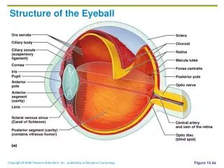
Anatomy of the Eyeball. Cavities of the Eyeball. separated by the lens anterior posterior. Anterior Cavity. between lens and cornea filled with aqueous humor (watery fluid) Divided into: - anterior chamber (anterior to iris) - posterior chamber (posterior to iris). Posterior Cavity.

E N D
Cavities of the Eyeball separated by the lens • anterior • posterior
Anterior Cavity • between lens and cornea • filled with aqueous humor (watery fluid) • Divided into: - anterior chamber (anterior to iris) - posterior chamber (posterior to iris)
Posterior Cavity • between lens and retina • filled with vitreous body (jelly-like substance) • also called vitreous chamber
Lens • elastic, transparent, biconcave structure • separates anterior and posterior cavities of eyeball • suspended from ciliary body by suspensory ligaments • tension on suspensory ligaments controls shape of lens
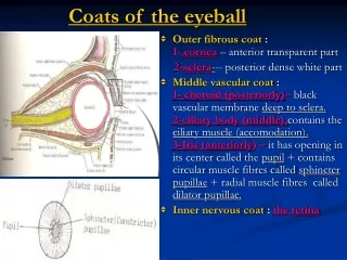
Layers of Eyeball (tunics) • fibrous - outer - sclera - cornea • vascular - middle - choroid - ciliary body - iris • retina - inner nervous
Layers of Eyeball iris sclera cornea pupil choroid ciliary body retina
Sclera - outer fibrous • “white of the eye” • outermost • protects eye • thick, tough connective tissue • capsule that maintains shape of eye • serves as point of attachment for extrinsic muscles • makes up 5/6 of sclera
Cornea - outer fibrous • anterior 1/6 of fibrous tunic; continuous with sclera • bulges forward, forming convex surface - refracts light rays as they enters eye • transparent - allows light rays to pass • lacks blood vessels • receives nutrition from lymph • has five layers
Cornea - outer fibrous (cont.) • has touch and pain receptors • injury causes scarring • most exposed part of eye • great ability to repair itself • only tissue that can be transplanted from person to person without rejection
Scleral Venous Sinus • also called canal of Schlemm • junction of sclera and cornea • drains aqueous humor from eyeball
Choroid - middle vascular layer • vascular layer; blood rich • contains dark pigment produced by melanocytes - absorbs pigment and prevents scatter of light after it passes through retina • anterior portion becomesciliary body and iris
Ciliary Body - middle vascular layer • thickest part of vascular tunic • forms internal ring in anterior part of eyeball • within are projections or folds called ciliary muscles - secrete aqueous humor into anterior cavity • lens is attached via suspensory ligaments
Iris - middle vascular layer • extends out from ciliary body • anterior to lens • thin diaphragm of connective tissue • seen from outside as colored portion of eye • has rounded opening called pupil • regulates amount of light entering posterior cavity of eyeball through pupil
Pupils • bright light or close up - pupils constrict • dim light or distance - pupils dilate
suspensory ligaments anterior chamber lens cornea posterior chamber pupil iris
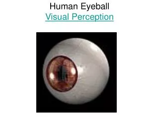
Retina - innernervous layer • light sensitive • is where light rays form an image • image travels via optic nerve to cerebral cortex • if image is not focused correctly, corrective glasses or lenses are required • contains photoreceptors - rods and cones
Photoreceptors • rods - 20 million - recognize gray tones and dim light • cones - 6 million - recognize primary colors • together they interpret intermediary colors • in moonlight only rods are functioning; therefore we cannot see colors
Fovea Centralis • depressed area in center of macula lutea - yellowish spot just lateral to optic axis of eyeball • has highest concentration of cones in retina • produces sharpest vision and best color perception
Optic Disc • also called blind spot • medial to optic axis • fibers from ganglion cells exit eyeball to form optic nerve • no photoreceptors; light striking this area produces no image
Color Blindness • inability to distinguish colors • caused by a lack or deficiency in one of the three cone photopigments • most common type is red-green color blindness • inherited condition affecting males more often than females - sex-linked
Intraocular Pressure • caused when scleral venous sinus is obstructed and reabsorption of aqueous humor cannot keep up with its secretion • pressure in chambers pushes lens back and puts pressure on vitreous body which in turn presses on retina which obstructs blood supply
Intraocular Pressure (cont.) • retinal cells die and optic nerve may atrophy causing blindness (glaucoma) • symptoms usually go unnoticed until damage is irreversible • disease can be detected by use of tonometer used to measure intraocular pressure
Glaucoma • group of eye diseases • characterized by an increase in intraocular pressure • pressure causes pathological changes in optic disk and visual field defects
Abnormal Flow of Intraocular Fluid (most common type) • egress is partially blocked causing increased accumulation of fluid causing increase pressure and eventual blindness
Acute Closed-Angle Abnormal Flow of Intraocular Fluid • egress is totally blocked causing permanent blindness suddenly
Accessory Structures • eyelids • lacrimal apparatus • extrinsic muscles • cranial nerves
Eyelid • Composed of: - skin covers outer surface - conjunctiva covers inner surface of eyelid and anterior surface of eyeball (except cornea)
Eyelid (cont.) • Composed of: - tarsal glands modified sebaceous gland (oil) open at edge of each eyelid also called Meibomian glands - muscles orbicularis oculi - surrounds eye levator palpebrae - in upper eyelid
Lacrimal Apparatus • lacrimal gland • superior and inferior canaliculi • lacrimal sac • nasolacrimal duct
Lacrimal Gland • located in upper portion of each orbit • secretes constant flow oftears - wash anterior surface of eyeball - maintain moist and clean environment for cornea and conjunctiva - contain antibacterial enzyme lysozyme that helps prevent eye infections
Superior and Inferior Canaliculi • collect tears after they have washed over eyeball Lacrimal Sac • collects tears from canaliculi Nasolacrimal Duct • connects lacrimal sac to nasal cavity where tears are swallowed
Extrinsic Muscles • arise from bones of the orbit • inserted into broad tendons on sclera • Six extrinsic eyeball muscles: lateral rectus medial rectus superior rectus inferior oblique inferior rectus superior oblique
superior oblique superior rectus trochlea inferior oblique lateral rectus (cut) medial rectus inferior rectus
Primary Actions of the Eye Muscles • abduction • adduction • elevation • depression
Abduction • contraction of lateral rectus moves pupil away from nose Adduction • contraction of medial rectus moves pupil towards nose
Elevation • contraction of superior rectus or inferior oblique muscles moves pupil upward Depression • contraction of inferior rectus or superior oblique muscles moves the pupil downward
Cranial Nerves of Eyeball Innervation • oculomotor (III) - branches innervate superior rectus, medial rectus, inferior oblique, and inferior rectus • trochlear (IV) - innervates superior oblique • abducens (VI) - innervates the lateral rectus