Download
1 / 13
130 likes | 220 Views
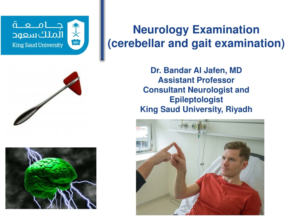
Neurology Examination (cerebellar and gait examination) Dr. Bandar Al Jafen , MD Assistant Professor Consultant Neurologist and Epileptologist King Saud University, Riyadh. CO-ORDINATION. WHAT TO DO Arms Finger-nose test Dysdiadochokinesis Legs Heel – shin test.

E N D
Neurology Examination (cerebellar and gait examination) Dr. Bandar Al Jafen, MD Assistant Professor Consultant Neurologist and Epileptologist King Saud University, Riyadh
CO-ORDINATION WHAT TO DO Arms Finger-nose test Dysdiadochokinesis Legs Heel – shin test The finger –nose test The heel-shin test
Cerebellar Examination andCoordination Hemisphere Dysfunction • Dysmetria on Finger-Nose-Finger Testing* • Irregularly-Irregular Tapping Rhythm* • Dysdiadochokinesis* • Hypotonia* • Impaired Heel-Knee-Shin* • Falls to Side of Lesion* • Nystagmus (Variable Directions) * All Deficits are Ipsilateral to the side of the lesion
Midline Dysfunction • Truncal Ataxia • Titubation • Ataxic Speech • Gait Ataxia • Acute Ataxia (unsteady Gait) • Chronic Ataxia (wide-based, steady Gait)
WHAT IT MEANS • Unilateral inco-ordination – ipsilateral cerebellar syndrome. • Bilateral inco-ordination – bilateral cerebellar syndrome. • Truncal ataxia, gait ataxia, without limb inco-ordination – midline cerebellar syndrome. • Unilateral cerellar syndrome – common causes: demyelination, vascular disease; rare causes: trauma, tumour or abscess. • Bilateral cerebellar syndrome – common causes: drugs (anti-convulsants), alcohol, demyelination, vascular disease; rare causes: hereditary cerebellar degenerations, paraneoplastic disorders, hypothyroidism. • Midline cerebellar syndrome: lesion of the cerebellar vermis – causes as for bilateral cerebellar syndrome.
The Neurological ExaminationGait Observe Different Aspects of Gait • Arm Swing • Base of Gait • Heel Strike • Time Spent on Each Leg • Posture of Trunk • Toe Walking • Heel Walking • Tandem Walking
GAIT • Always examine patient's gait. It is a co-ordinated action requiring integration of sensory and motor functions. The gait may be the only abnormality on examination. The most commonly seen are: hemiplegic, parkinsonian, ataxic and unsteady gaits. • Romberg's test is conveniently performed after examination the gait. This is a simple test primarily of joint position sense. • Ask the patient to walk • Ensure you are able to see the arms and legs adeguately. • Is the gait symmetrical? • Gait can usually be divided into symmetrical and asymmetrical even though the symmetry is not perfect. • If symmetrical: • Look at the size of paces • Small or normal? • If small paces: • Look at the posture and arm swing • Stooped with reduced armswing – parkinsonian(may be difficult to start and stop –festinant). • Upright with marked armswing – marche a' petits pas.
If normal paces: • Look at the lateral distance between the feet • normal • widely separated - broad based • Legs unco-ordinated – cerebellar • Crossing over, toes dragged – scissoring. • Look at the knees • normal • knees lifted high – high-stepping. • Look at the pelvis and shoulders • normal • marked rotation of pelvis and shoulder –waddling. • Look at the whole movement • normal • disjointed as if forgotten how to walk, patient frequently appears rooted to pot – apraxic. • bizarre, elaborate and inconsistent – functional. • If asymmetrical • Is the patient in pain? • yes – painful or antalgic gait. • Look for a bony deformity • orthopaedic gait.
Does one leg swing out to the side? • yes – hemiplegic gait. • Look at the knee heights • normal • one knee lifts higher – foot drop. • Ask the patient to walk as if on a tight – rope (demonstrate) • if patient fall consistently – unsteady • may fall predominantly to one side. • Ask the patient to walk on his heels (demonstrate) • If unable to – foot drop. • Ask the patient to walk on his toes (demonstrate) • If unable - weakness of gastrocnemius. • Parkinsonian: indicates basal ganglion dysfunction – common causes: Parkinson's disease, major tranquillisers. • Scissoring: indicates spastic paraparesis – common causes: cerebral palsy, multiple scelrosis, cord compression. • Sensory ataxia: indicates loss of joint position sense (Romberg's positive) – common causes: peripheral neuropathy, posterior column loss (see below). • Cerebellar ataxia: veers towards side of lesion – common causes: drugs (e.g. phenytoin), alcohol, multiple sclerosis, cerebrovascular disease.
Waddling gait: indicates weak or ineffective proximal muscles – common causes: proximal myopathies, bilateral cogenital dislocation of the hip. • Apraxic gait: indicates the cortical integration of the movement is abnormal, usually with frontal lobe pathology – common causes:normal pressure hydrocephalus, cerebrovascular disease. • Hemiplegic: unilateral upper motor neurone lesion – common causes: stroke, multiple sclerosis. • Foot drop: common causes – unilateral: common peroneal palsy, pyramidal lesion, L5 radiculopathy. Bilateral: peripheral neuropathy. • Non-neurological gaits • Painful gait:common causes:arthritis, trauma – usually obvious. • Orthopaedic gait: common causes: shortened limb, previous hip surgery, trauma.