Understanding Contrast Radiography Techniques
450 likes | 898 Views
Learn about contrast radiography, essential methods, contrast media types, advantages, and ideal contrast mediums. Discover common uses and examples of contrast radiography in medical imaging procedures. Explore principles and interpretation of contrast studies in the gastrointestinal tract.

Understanding Contrast Radiography Techniques
E N D
Presentation Transcript
Basic Radiographic Opacities • Radiographic image: It is produced when x- ray goes through the body part: penetration and absorption, hence== What you got?? • Basic radiographic opacities Air Fat Water Bone Metal/+Contrast BLACKGREY GREY GREY WHITE
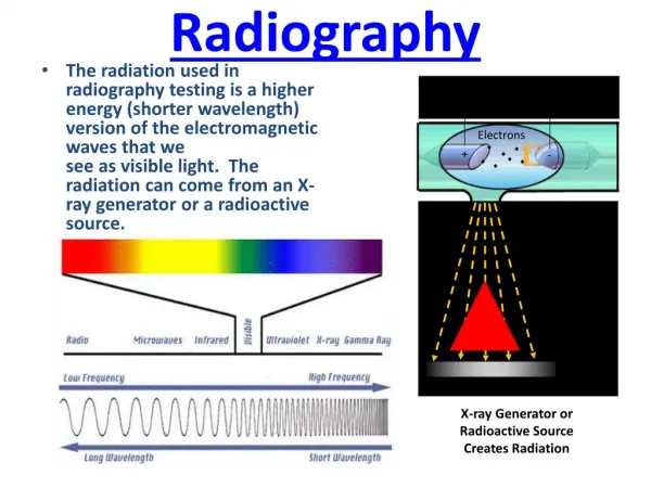
Contrast Radiography Contrast radiographic techniques are used to identify a soft tissue structure or an organ which may be difficult or impossible to visualize clearly in plain films due lacking of contrast with surrounding tissue. Contrast radiographic techniques employ the use of contrast agents like Barium Sulphate, Iodides, Air etc. The contrast may be a positive contrast barium or negative contrast like air. Tissue radio density and its surrounding is deliberately altered, for better visualization and demarcation.Group of radiographic procedures performed by administration of a contrast medium • What for?? • Visualization of individual organs • Enhance lesions in a particular organ • Some physiologic information • Always performed after a survey radiograph
Advantages: • Structures or organs can be evaluated more effectively for their size, shape and positions. • Valuable information can be gained regarding serosal and mucosal surfaces of hollow organs. • In some instances some idea of the function of the organ can be formed Ideal Contrast Medium: • Any contrast medium used should have following qualities. • It should have desired Z number. • It should be inert or its metabolic byproducts should not be toxic. • It should be retained in the area of interest only for the desired periods.
Contrast Media • Positive Contrast Media : Those materials, which increase radio density of the structure or tissue in relation to surrounding tissue, are called positive contrast media. (absorb X rays = radiopaque) • Barium (inert, not metabolized or absorbed) • Liquid • Paste • Iodine: Tri – iodinated derivatives of benzoic acid • Ionic • Non – ionic • Cholecystopaques • Viscous and oily agents.
Negative Contrast Media : Those materials, which relatively decrease the radio density, are negative contrast media. (do not absorb X rays = radiolucent) • Air • Carbon dioxide • Nitrous oxide
Barium sulphate is exclusively used for outlining alimentary tract. It is insoluble and not absorbed in the body. Therefore its use should be avoided if perforations are suspected. It is available as powder, suspension or paste. Not used with ruptured GIT as it will lead to inflammation, formation of granulomatus mass and fibroma. Barium Sulphate preparation • Insoluble • Non absorbable by GIT
Water Soluble Iodine Compound Sodium salt of Iothalamic Meglumine salt of Iothalamic Sodium salt of Ditriazote Meglumine salt of Ditriazote Commonly used contrast medium having low osmolarity. These form the largest single group of contrast media. These are commonly used for outlining the urinary system. Commonest conventional agents are : These are quickly eliminated by the system.
Water soluble organic iodine.Very useful in liver functionsExcreted exclusively through biliary-system Hence used for outlining biliary system and gall bladderMostly used as IV CHOLECYSTAPAQUES
Viscous and Oily preparation Used for Myelography if Non-iodine low osmolarity medium not available. These agents are less irritant because of their immiscibility with water, there are not suitable for intravascular use. Their use is limited to lymphangiography, dacrocystorhinography, tracheography, bronchography, and hysterosalphangiography.
EXAMPLES OF CONTRAST RADIOGRAPHY Dacrocystorhinography : Nasolacrymal duct Sialography : Salivary gland Bronchography : Bronchioles Reticulography : Reticulum Pneumocystography : Abdominal Cavity Intravenous Pyelography : Urinary tract Myelography : Spinal Cord Arthrography : Joints Fasciography : Tendon and associated structures Osteomedullography : Channels of long bones. Angiography : Arteries Urethrography : Urethra Cystography : Urinary Bladder
A B C submucosal or intramural mass extrinsic mass mucosal mass Principles
A B Mucosal Mass
MASS Extrinsic lesions
Contrast studies for the GI tract • Esophagogram, esophagram, esophogram • Indications: • Suspected esophageal disease • Confirm questionable findings on survey rads • Esophageal motility • Procedure • Survey rads • Sedation not recommended (Acepromazine) • Interpretation • No distention, you can see the bolus • Longitudinal folds and herringbone pattern (cats)
Gastrography • Indications • Suspected gastric wall lesion • Gastric location • Pyloric obstruction (check on fluoroscopy) • Procedure • Double contrast study (barium + air) • Good for gastric mucosa • Single contrast (either barium or air-pneumogastrogram) • Barium: good for gastric motility • Air: good for foreign body • Interpretation • Position • Rugal folds • Motility (gastric emptying) (follow with fluoroscopy)
Contrast Studies of Intestine • Indications • Size, shape, mucosa of SI • Suspect of SI disease • Diagnosis of obstruction • Contraindication • Barium if suspected rupture of hollow viscous, use iodine (ionic) instead • Procedure • Preparation • Interpretation • Lumen diameter • Mucosa
Contrast Studies for the Urinary Tract • Excretory Urography (EU) – Intravenous Pyelogram (IVP) – Intravenous Urography (IVU) • Indications • Evaluate kidney morphology – position. Ureters. • Suspected renal disease • Increased radiopacity in the retroperitoneal space • Gross idea of kidney function • Procedure • Preparation required (24 h fasting; enema) • IV access/dose • Radiographic views
Excretory Urography (EU) – Intravenous Pyelogram (cont’d) • Interpretation • Vascular phase • 5 – 10 sec • Nephrogram phase • 1 – 2 min • Normal pattern: Initial=Good, then=gradual decrease • Abnormal patterns: • Poor – persistent • Poor – continued increase/ decrease • Good – continued increase
Cystography • Indications • Suspected bladder disease • Caudal abdominal mass • Bladder rupture • Persistent dysuria /hematuria /stranguria • Congenital anomalies • Procedure • 24 hour fast – enema • Empty bladder • Four views • Positive contrast/ Double contrast • Negative contrast (pneumocystogram)
Cystography • Interpretation
Negative contrast cystographyCatheterise the bladder (using sterile technique)Drain all the urine Instill air or gas into the bladder until it becomes slightly turgid (judged by palpation of the bladder through the abdominal wall)Take lateral and ventrodorsal radiographs Retrograde cystography
Double contrast cystography The 3rd contrast procedure is to drain positive contrast and re-instill air. Radiographs reveal a double contrast study of the bladder. Any positive contrast which adheres to the mucosa is because of inflammatory bladder wall lesions. Also help to identify small calculi which were previously not visible on the negative or positive contrast studies. Polyps, bladder wall infiltrating masses, urachal diverticuli and inflammatory bladder wall thickenings will all be identified using this sequence of bladder studies.
Bladder images demonstrating a mass infiltrating the ventral bladder wall. Negative Double contrast
Urethrography • Indications • Investigate stranguria / hematuria / dysuria • Urethral rupture • Malformations • Procedure • Positive contrast medium (iodine) • Vaginourethrography • Interpretation
Contrast radiography of genital tract Substance should be water soluble to permit its absorption and elimination from the uterus And also comparatively viscous so that it does not easily spill back from the uterus. The amount required will vary with the size and nature of the uterine condition, and it is usual to introduce 1 to 6 ml (depending on the size of the dog) and to administer additional amounts if radiography indicates that this is required.
Pneumoperitonium Air or oxygen is employed. The amount required is not very precise and varies from 200 to 1000 ml, depending on the size of dog.