Download
1 / 54
550 likes | 1.06k Views
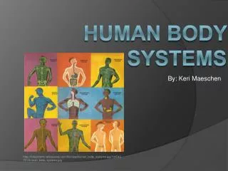
By: Keri Maeschen. Human body systems. http://finleymslmc.wikispaces.com/file/view/human_body_systems.jpg/106542751/human_body_systems.jpg. Table of Contents. Digestive……………………………3-12 Circulatory…………………...........13-26 Respiratory…………………………27-35 Immune……………………….........36-46

E N D
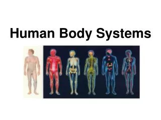
By: Keri Maeschen Human body systems http://finleymslmc.wikispaces.com/file/view/human_body_systems.jpg/106542751/human_body_systems.jpg
Table of Contents • Digestive……………………………3-12 • Circulatory…………………...........13-26 • Respiratory…………………………27-35 • Immune……………………….........36-46 • Excretory…………………………...47-54
Function • The function of this system is to transform food into a slightly different form to be allowed into the next compartment. Food starts as complex particles and is changed into basic elements that are absorbed by the body, while the waste is expelled through the anus.
Organs • Salivary Glands: produces saliva which is a lubricant for chewing and swallowing food (accessory) • Mouth: place where food enters mouth and chewing takes place (alimentary) • Pharynx: aids in moving food from the mouth to the esophagus and is where swallowing takes place (alimentary) • Esophagus: Delivers food from the pharynx to the stomach by peristalsis and the lower esophageal sphincter keeps food from going backwards (alimentary) • Stomach: holds, mixes, and grinds food; secretes acids and enzymes to breakdown food (alimentary) • Small Intestine: Uses enzymes from the pancreas and bile from the liver; peristalsis moves food from duodenum (breaks it down), then jejunum, and ileum (both absorb nutrients) (alimentary) • Liver: Makes and secretes bile, and cleanses and purifies blood (accessory) • Pancreas: secretes enzymes into the small intestine where enzymes are made to break down fat, protein, and carbohydrates from food (accessory) • Gall Bladder: during meals, it contracts and sends stored bile to the small intestine (accessory) • Large Intestine: (colon) waste product is passed through by peristalsis and is stored until it needs to be emptied. It includes the cecum which starts near the appendix, then the ascending colon which is above the cecum, next is transverse colon which is across the top, then the descending which is on the opposite side going up and down, and the sigmoid colon which is the end. (alimentary) • Appendix: unknown function; may be for storing good bacteria (accessory) • Rectum: receives waste from colon and signals that it is ready to be evacuated (alimentary) • Anus: prevents waste from coming out when it isn’t supposed to (alimentary) • Sphincters: circular muscle that constricts or relaxes when food is passed through (accessory)
Digestion of Large Molecules • Large particles are digested first by placing them in the mouth. The large particles are chewed using the teeth and the mouth, along with saliva to lubricate it and break it down with the enzyme amylase. Next, the food is in a smaller form and is swallowed by passing through the pharynx and the esophagus to the stomach. In the stomach, it is stored, ground up, and hydrochloric acid mixes with the food to create chyme. After that, chyme enters the duodenum of the small intestine where enzymes from the gall bladder, pancreas, and liver are secreted that further break down the food into simpler elements. Villi in the small intestine allow for absorption of the elements. Next, the liquefied chyme enters the large intestine, where it becomes waste and as it passes through, water is absorbed and it is turned into a solid. Waste is collected in the rectum where it is stored until there is a signal saying that it needs to be emptied. It is then eliminated through the anus. This process is essential because humans eat large molecules and they must be made small enough for our bodies to absorb.
Role of Enzymes • Enzymes are protein molecules that make life processes happen by acting as change agents in the chemical reactions that take place in the body. They build up or break down substances needed. Amylase is found in saliva and it breaks down carbohydrates. Pepsin is found in the stomach and is a protease enzyme that breaks down food. Small intestine enzymes include lactase (which breaks down milk), DPP IV (which breaks down proteins), and disaccharides (which break down sugars and starches). Basically, the role of enzymes is to break down the foods we eat so they are able to be absorbed into the blood stream.
Physical vs. Chemical Digestion • Physical digestion is when digestion takes place without the aid of chemicals. Chewing and mixing are forms of physical digestion. • Chemical digestion is when digestion takes place with enzymes catalyzed. Polysaccharides to monosaccharides, proteins to amino acids, and fats to glycerol + amino fatty acids. • Both are used to break down foods
Carbohydrate, Protein, & Lipid Digestion • Carbohydrates (starch and sugar) are digested by amylase and are then turned into glucose. This takes place in the mouth. • Proteins are digested by proteases. These include pepsin (in the stomach), and trypsin (in the small intestine). • Lipids (fats) are not water soluble, so they are covered in bile. Lipase is the enzyme that digests fat and is located in the digestive tract.
Disorders • Lactose Intolerance: This happens when the body lacks the enzyme lactase, which is needed to digest lactose. Both children and adults are affected by this disorder. It can be caused by diseases or injuries to the small intestine and symptoms include bloating, cramps, gas, diarrhea, and nausea and as larger amounts of lactose are taken in, symptoms get worse. Between 30 and 50 million Americans have this and can be treated by eating less dairy products, or even eliminating them from a diet. • Stomach Ulcers: Ulcers are open lesions or sores and can be found in the skin or mucous membranes of the body. A stomach ulcer can also be known as a gastric ulcer. Lifestyle, stress, and diet may aid in formation of the ulcers, but aren’t the main cause. Hydrochloric acid and pepsin are the main causes and contribute to ulcer formation. Symptoms include a feeling of gnawing or burning in the stomach. About 6,000 Americans die from ulcers or ulcer-related problems. This can be treated by lowering acid amounts in the stomach.
Sources • http://catalog.nucleusinc.com/imagescooked/1874W.jpg • Campbell & Reece AP Biology Book • http://www.webmd.com/heartburn-gerd/your-digestive-system • http://www.lrn.org/Content/Lessons/digestive.html • http://www.sparknotes.com/health/digestion/section2.rhtml • http://www.earthyfamily.com/A-enzymesDig.htm • http://www.enzymestuff.com/digestion.htm • http://www.indiana.edu/~nimsmsf/P215/p215notes/PPlectures/Printables/Digestion.pdf • http://socialmediasystems.com/rothmanguide/files/2009/11/837375_mouth.jpg • http://www.nlm.nih.gov/medlineplus/ency/images/ency/fullsize/8945.jpg • http://digestion.ygoy.com/the-5-stages-of-digestion/ • http://www.healthcentral.com/common/images/1/1055_3037_5.jpg • http://www.scientificamerican.com/media/inline/8F0B3BA6-A03E-CF2D-A538AB82900B1318_1.jpg
Function • The circulatory system transports materials (including gas, nutrients, wastes, and hormones), contains cells that fight infection, stabilize ionic concentration and pH of fluids the body, and transports heat to maintain temperature.
Blood Vessels • Arteries: • They have a thick wall for high blood pressure. • There is a thick outer layer of collagen and elastic fibers. • They have thick layers of muscular, circular, and elastic fibers. • The blood goes from the heart to the small intestine. • Contains a narrow lumen to maintain high pressure. • Veins: • They have thin layers with a few muscular, circular, and elastic fibers. • Muscles squeeze them because of their thin walls. • Contains thin outer layer of collagen and elastic fibers. • There is a wide lumen for slow blood flow. • Valves allow the blood to stay and not flow back. • Capillaries: • They connect veins and arteries together. • Capillaries are thin and moist for diffusion. • They are narrow, but allow large quantities. • Pores allow diffusion of phagocytes and plasma.
Blood Route • Oxygenated blood from the lungs travels to the left side of the heart via pulmonary veins, and empties in the left atrium. From there it goes to the left atrioventular (bicuspid) valve and to the left ventricle. Seventy percent of this flow comes in when the heart is relaxed, and the other thirty percent comes in when the atrium contracts. There is a slight delay before the ventricle contracts, and blood is forced to exit to the aorta. The atrioventular valve closes to prevent backflow, and the aorta is closed off by the aortic semilunar valve which snaps shut only when backflow tries to take place. From the aorta, blood follows vessels throughout the body (other than the lungs) which makes up the systemic circulation. It takes blood to organs, and gives the oxygen to body tissues, while taking carbon dioxide from them. After flowing through the capillaries, the blood goes through the heart again, except through the right side. The superior vena cava drains the top, while the inferior vena cava drains the bottom and both dump it into the right atrium. Next, it goes through the right atrioventricular (tricuspid) valve to the right ventricle. Then it passes through the pulmonary semilunar valve into a single pulmonary artery, which branches into arteries that carry deoxygenated blood to the lungs. Next, it returns from the lungs to the left side of the heart replenished with oxygen and the process repeats.
Blood Composition • Blood is a liquid tissue that is made of plasma, erythrocytes, leukocytes, and platelets. • Plasma is the straw-colored liquid that contains the cells. • Erythrocytes, or red blood cells, make up ninety-nine percent of the cells in blood. Two types of protein (A and B) are found there and different combinations make up the four blood types (A, B, AB, O) • Leukocytes, or white blood cells, are a large source of defense for blood and “clean” it by picking up dead cells. These cells can be used to diagnosis sicknesses. • Platelets are used when damage takes place on the body. They trap the open wound and clot together to form a scab to prevent dangerous loss of blood
Erythrocytes • Erythrocytes are flat and cylindrical, which allows more oxygen and carbon dioxide to be carried throughout the body. They are also very flexible and move efficiently through veins and vessels. They are made of hemoglobin which is composed of iron and proteins.
Open vs. Closed Circulatory Systems • Open: Circulatory fluid, or hemolymph, bathes organs directly. The heart contracts pumping the hemolymph through the circulatory vessels into interconnected sinuses. In the sinuses, chemical exchange occurs between the hemolymph and body cells. The heart relaxes and draws hemolymph back in through pores, and body movements help circulate hemolymph by squeezing sinuses. An example is lobsters and other arthropods and mollusks. • Closed: Blood is confined to vessels and is distinct form the interstitial fluid, unlike the open system. One or more heart pumps blood into vessels that branch off into smaller ones through the organs. Materials are exchanged between the small vessels and the interstitial fluid bathing the cells. Earthworms are an example.
Animal Circulation: Fish • Fish have single circulation where blood passes through the heart once in a complete circuit. Contraction of the ventricle pumps blood to the gills where there is a net diffusion of oxygen into the blood and carbon dioxide out of the blood. When blood leaves the gills, capillaries converge into a vessel that carries oxygen right blood to capillary beds through the body, then blood returns to the heart.
Animal Circulation: Amphibians • Amphibians have a three-chambered heart with two circuits of blood flow including the pulmocutaneous and systemic. They have a heart with three chambers (two atria and one ventricle). When underwater, circulation is adjusted and for the most part blood flow is shut off due to ineffective lungs temporarily, but blood flow continues to the skin which is where gas exchange takes place.
Animal Circulation: Reptiles • Reptiles have three chambered hearts and a septum that partially divides the ventricle into right and left chambers. In some animals (crocodiles, caimans, and alligators), the septum is complete, but the pulmonary systemic circuits are connected where the arteries exit the heart.
Animal Circulation: Mammals and Birds • Mammals and birds have a four-chambered heart, but birds have slightly different major vessels near the heart. The ventricle is completely divided so there are two atria and two ventricles. The left side receives and pumps oxygen-rich blood, while the left side receives and pump oxygen-poor blood.
Disorders • Arteriosclerosis happens when there is an accumulation of fatty acids in the arteries, which thickens and stiffens the walls. This makes blood unable to flow and may cause blood clots, which may lead to heart attack which is one of the symptoms. This can be treated by eating less fatty foods, exercising, and limiting alcohol intake. In America, for sixty-five percent of men and forty-seven percent of women, the first symptom is heart attack or sudden cardiac death. • Hypertension, also known as high blood pressure and the pressure that is high is about 140/90. Symptoms include confusion, fatigue, headache, nosebleed, and vision change. Approximately 927 million people worldwide suffer from this problem. This can be treated by taking alpha or beta blockers, exercising, eating healthy, using diuretics, or using vasodilators.
Sources • http://www.ashlandschools.org/morgan_cottle/body/circ.gif • Campbell & Reece AP Biology Book • http://faculty.clintoncc.suny.edu/faculty/michael.gregory/files/bio%20102/bio%20102%20lectures/circulatory%20system/circulat.htm • http://www.phschool.com/science/biology_place/biocoach/cardio2/vessel.html • http://library.thinkquest.org/28807/data/circ3.htm • http://www.chelationtherapyonline.com/articles/images/30_3c.jpg • http://image.wistatutor.com/content/transportation/single-blood-circulation-fish.jpeg • http://brucemhood.files.wordpress.com/2008/09/blood_cells.jpg • http://www.sumanasinc.com/webcontent/animations/content/human_heart.html • http://en.wikibooks.org/wiki/IB_Biology/Human_Health_and_Physiology • http://simscience.org/membranes/advanced/essay/blood_comp_and_func1.html • http://www.ncbi.nlm.nih.gov/pubmedhealth/PMH0001502/ • http://www.cosmosmagazine.com/files/imagecache/news/files/20070416_bloodpressure.jpg • http://library.thinkquest.org/5777/images/circulatory.gif
Function • The function of this system is to take in oxygen by inhalation and take the oxygen to the cells throughout the body, while carbon dioxide is taken out through exhalation. Besides respiration, the respiratory system also aids in coughing and vocals.
Alveoli • Alveoli are grouped together like grapes. • Oxygen inhaled diffuses through the walls of the alveoli and goes into red blood cells. • They have thin walls for efficient gas exchange. • The large surface area compared to volume allows more intake of gas. • They are lined with fluid for gas to dissolve. • Alveoli are surrounded by many capillaries
Transports • Carbon dioxide is transported in blood by a few processes. It diffuses along the concentration gradient into the plasma through the tissues. When it is in the blood most of it enters the red blood cells, some stays in the plasma. • Oxygen is transported in blood in plasma as dissolved oxygen and as a chemical combination with hemoglobin.
Trace Oxygen • First, air is taken in by either the mouth or nose. • Next, oxygen passes through the larynx and trachea. • Trachea splits into two bronchi and those divide into bronchial tubes where oxygen passes through after the larynx and trachea. • Bronchial tubes connect to the lungs and the bronchial tubes become smaller tubes that connect to alveoli, or tiny sacs where the gas is exchanged. • Capillaries bring in the carbon dioxide from the heart and diffuse the oxygen into the hemoglobin of the blood to take back to the heart.
Breathe In, Breathe Out • Inhalation takes place when the diaphragm and intercostals muscles contract. The diaphragm moves down while the intercostals push the rib cage up. This reduces the air pressure to below atmospheric pressure, which allows air to rush in. The volume of the thoracic cavity is also increased. • Exhalation takes place when the diaphragm and intercostals relax and the diaphragm moves up while the intercostals move down. The thoracic cavity volume decreases and pressure is above atmospheric pressure, causing air to release out of the airways.
Disorders • Asthma: inflammatory disorder of the airways where the muscles become tight and air passages swell. Symptoms include wheezing, shortness of breath, and coughing. Approximately 1 in 15 Americans suffer from asthma. Treatment includes inhalant corticosteroids and inhibitors. • Pneumonia: is an infection in the lungs where bacteria or viruses infect. Symptoms include coughing, shaking, chills, and shortness of breath. About 4.8 million people in America have had pneumonia. This can be treated by amoxicillin, Avelox, or other antibiotics.
Sources • http://respiratorysystemdiagram.net/respiratorydetail.gif • Campbell & Reece AP Biology Book • http://www.buzzle.com/articles/respiratory-system-functions.html • http://www.curoservice.com/parents_visitors/lungs_circulation/structure_alveoli.asp# • www.mpoullis.net/.../Transport%20of%20O2%5B1%5D.CO2.doc • http://www.ambulancetechnicianstudy.co.uk/respsystem.html • http://www.aafa.org/display.cfm?id=9&sub=42#prev • http://www.ncsl.org/Portals/1/oldsite/programs/environ/envhealth/asthmarates-99.gif • http://www.ncbi.nlm.nih.gov/pubmedhealth/PMH0001200/ • http://www.empowher.com/files/ebsco/images/pneumonia%20lung%20fluid.jpg
Function • The immune system is a network of organs, tissues, and chemicals made up to protect the body from things like infection or sickness. Parts that play a role in this system include the lymph nodes, spleen, bone marrow, thymus gland, and tonsils.
Organs • Thymus produces T cells and puts them in the blood stream. First, immature T cells derive from bone marrow and go through a maturation process once they get to the thymus. Beneficial ones are spared while harmful ones are evoked. • Spleen is made up of B cells, T cells, macrophages, dendrite cells, natural killer cells, and red blood cells. Its purpose is to filter the blood by collecting antigens that pass through. • Lymph nodes filter the bodily fluid called lymph and is made mostly of T cells, B cells, dendrite cells, and macrophages.
Pathogens, Antigens, & Antibodies • When humans have a virus once, they probably won’t get it again because the immune system is able to remember the virus. On antibodies, they have a bonding site to recognize antigens, which have unique markers making them easy to identify. The antibody tags the antigen which triggers mechanisms to destroy it. Some mechanisms include when it is bound to the antibody it stops viruses from attaching to a host cell or pathogens are clumped together for phagocytes to destroy.
Innate vs. Acquired • Innate immune system is the system that you are born with and protects against antigens. They are the first line of defense in the immune system because they create a barrier to keep harmful materials away. Examples include cough reflex, tear enzymes, stomach acid, skin, and mucous. • Acquired immunitydevelops when there is exposure to some antigens. Once your system has had a sickness, it is easier for your body to fight it off, which means a system is built to defend against that specific antigen. An example is getting the chicken pox, but you do not get them again because your body recognizes it. • Both defend our bodies against sicknesses and disorders that may affect us.
Active vs. Passive • Active immunity is when antibodies are developed after an antigen stimulates it. In response to infection, clones of memory cells form. An example is immunization or vaccination like using cowpox vaccination. • Passive immunity involves antibodies developed from another body. Examples include young infants (have them from their mother), and antiserum which is from another person or animal. • Both can be induced artificially. They are both ways to protect the body.
Humoral vs. Cell-mediated • Humoral uses antibodies that are used to target cells that are potentially dangerous. • Cell-mediated activates macrophages, natural killer cells, T-lymphocytes, and cytokines. • Both are fundamental adaptive systems.
B vs. T lymphocytes • B cells are involved in humeral immunity. They produce large quantities of antibodies which neutralize the bacteria or virus. • T cells are involved in cell-mediated immunity. Some produce cytokines that direct the other immune response but others produce cytotoxic T cells, which make toxic granules that has enzymes that induce death of infected cells.
Antibiotics & Bacteria • Bacteria must live on a host so they are easier to kill than viruses. Antibiotics can block metabolic pathways of bacteria, which inhibits cell wall formation and protein synthesis. This causes death. In order for a virus to be killed, the human cell must be killed also.
Disorders • HIV/AIDS: Human Immunodeficiency Virus is a virus similar to the flu, but our immune systems cannot get rid of it because they take over CD4 and T-cells to make copies of itself and destroy them. Acquired Immunodeficiency Syndrome is the final stage of HIV where people have badly infected immune systems. Many people do not have symptoms for a long time, but eventually feel like they have “a very bad flu”. 33.4 million people in the world are living with AIDS. People are treated with antiretroviral therapy which prevents HIV from growing, although it cannot be completely cured. • Sarcoidosis: This is a disease where the lymph nodes, lungs, liver, eyes, skin, or other tissues become inflamed. Symptoms include abnormal breathing, coughing, fatigue, rashes, seizures, enlarged liver, dry mouth, and many more. Approximately 1 in 5,000 people has Sarcoidosis. This disease eventually heals over time, but severe cases are treated by corticosteroids.
Sources • http://www.phoenix5.org/glossary/graphics/lymphatic_system.gif • Campbell & Reece AP Biology Book • http://www.immunesystemboost.net/functions.htm • http://www.thebody.com/content/art1788.html • http://myimmunehealing.com/images/Immune_System_Organs.jpg • http://www.umm.edu/ency/article/000821.htm • http://zesterandpeeler.com • http://users.rcn.com/jkimball.ma.ultranet/BiologyPages/B/B_and_Tcells.html • http://www.ncbi.nlm.nih.gov/pubmedhealth/PMH0001140/ • http://www.metrohealth.org/images/Patient%20Services/pulmonary/sarcoidplaque.jpg • http://www.web-books.com • http://www.antibioticsandalcohol.org/wp-content/uploads/2011/02/antibiotics-and-alcohol-drugs.jpg • http://en.wikipedia.org/wiki/Lymphocyte#T_cells_and_B_cells
Function • This system gets rid of wastes in the body and regulates the amount of water and ions present in fluids. Toxics are removed to prevent buildup and death.
Wastes • Ammonia: After the amino group is removed from amino acid, ammonia is formed and is highly soluble in water, but very toxic. Animals that excrete ammonia include bony fishes, aquatic invertebrates, and amphibians because it can be easily eliminated in water. • Urea: Urea is produced in the liver and requires more energy than ammonia to be made. Animals that excrete urea include terrestrial amphibians and mammals, because it is less toxic and can be concentrated to conserve water. • Uric Acid: Uric Acid is relatively insoluble and nontoxic, so it can accumulate in eggs without damaging the embryos and synthesis of uric acid requires more energy than urea synthesis. Insects, reptiles, birds, and some dogs secrete uric acid, and they do this because it is not very toxic and isn’t very soluble in water.