Download
1 / 1
10 likes | 100 Views
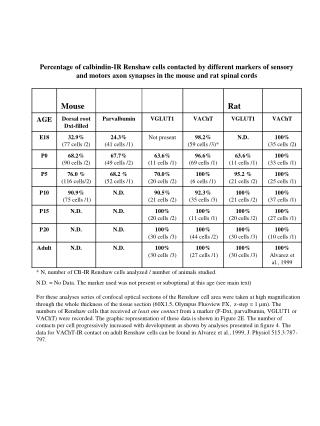
Percentage of calbindin-IR Renshaw cells contacted by different markers of sensory and motors axon synapses in the mouse and rat spinal cords. * N, number of CB-IR Renshaw cells analyzed / number of animals studied.

E N D
Percentage of calbindin-IR Renshaw cells contacted by different markers of sensory and motors axon synapses in the mouse and rat spinal cords * N, number of CB-IR Renshaw cells analyzed / number of animals studied. N.D. = No Data. The marker used was not present or suboptimal at this age (see main text) For these analyses series of confocal optical sections of the Renshaw cell area were taken at high magnification through the whole thickness of the tissue section (60X1.5, Olympus Fluoview FX, z-step = 1 µm). The numbers of Renshaw cells that received at least one contact from a marker (F-Dxt, parvalbumin, VGLUT1 or VAChT) were recorded. The graphic representation of these data is shown in Figure 2E. The number of contacts per cell progressively increased with development as shown by analyses presented in figure 4. The data for VAChT-IR contact on adult Renshaw cells can be found in Alvarez et al., 1999, J. Physiol 515.3:787-797.