Regulation of Apoptosis: Understanding Cellular Pathways
140 likes | 165 Views
Explore connections between metabolic pathways and apoptosis regulation. Learn about BH3-only proteins, IAPs, cell proliferation vs. apoptosis. Discover the role of IAP proteins in Drosophila and BCL-2 related proteins. Understand the intricate structures and regulatory mechanisms of apoptosis pathways.

Regulation of Apoptosis: Understanding Cellular Pathways
E N D
Presentation Transcript
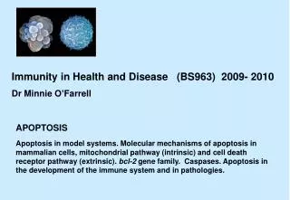
Apoptosis II • Connections between metabolic pathways and the regulation of apoptosis • BH3-only proteins as sensors • IAPs and decoy receptors • Proliferation vs. apoptosis in Myc signalling Sources: Helmreich, Gomperts, Voet and Voet, Danial and Korsmeyer (2004), “Cell Death: Critical Control Points.” Cell116, 205–219. Jiang and Wang (2004), “Cytochrome C-Mediated Apoptosis.” Annu. Rev. Biochem.73, 87-106. Hipfner and Cohen (2004), “Connecting proliferation and apoptosis in development and disease.” Nature Rev. Mol. Cell. Biol., 5, 805-815. Green and Kroemer (2004), “The Pathophysiology of Mitochondrial Cell Death.” Science, 305, 626-629. Jason Kahn: Apoptosis II
Regulation of Apoptosis • Recall intrinsic and extrinsic pathways: • Trimeric cell-surface receptors mediate extrinsic signalling, through initiator caspase-8 autoactivation (proteolysis) via localization • BCL-2 related proteins have proapoptotic or antiapoptotic effects through controlling the intrinsic pathway, in which cytochrome c release from mitochondria leads to apoptosome formation and caspase-9 activation. • These pathways are inhibited by, for example, decoy cell surface receptors (they lack cytoplasmic domains), cross-activated pathways that inhibit caspase activation, and by Inhibitors of Apoptosis (IAP’s) that block caspases. • Presumably this prevents the initiation of apoptosis by random clustering of a few receptors or other weak signals. When the switch is thrown, IAP’s are targets for caspases. Jason Kahn: Apoptosis II
IAP Proteins • The first IAP discovered was the baculovirus p35 protein, which inhibits host cell apoptosis during infection. Hence the critical protein domain of the IAP’s are called BIRs, for baculovirus IAP repeat • The BIR domains block access to the active sites of their target caspases: here the caspase-3 subunits are blue and the BIR2 domain is green. It binds Zn++ (purple) Active site Figure 34-111 X-Ray structure of caspase-7 in complex with the tetrapeptide aldehyde inhibitor Ac-DEVD-CHO Figure 34-120 X-Ray structure of caspase-3 in complex with the XIAP BIR2 domain and its N-terminal extension. Jason Kahn: Apoptosis II
Regulation of IAP’s • Smac/DIABLO (two names, one homodimeric protein), released from mitochondria along with cyctochrome c, binds the BIR domain of XIAP (X-linked IAP). This frees caspase-3 to proceed into apoptosis. • As always, confers irreversibility. • Illustrates feed-forward mechanism: increasing the ability to process a substrate at the same time as the level of that substrate is increased • Smac/DIABLO releases caspase-3Smac/DIABLO displaces caspase-9 from XIAPCaspase-3 cleaves off IAP-binding domain of caspase-9Thus the level and the processing of caspase-9 are both upregulated. • Degradation of IAP’s can be proapoptotic or antiapoptotic, depending on whether the cleaved product remains bound to the caspase; if it is, then both the IAP and the caspase can be targeted by the proteasome. Jason Kahn: Apoptosis II
IAPs in Drosophila • Different organisms have common mechanisms for apoptosis, but they vary in the relative importance of these mechanisms • Drosophila relies more heavily on IAP’s than we do • The RHG (Reaper, Hid, and Grim) proteins are regulated by upstream signals, and they in turn act by antagonizing the DIAP1 protein • Note regulation of RHG proteins and caspases by micro-RNAs Jason Kahn: Apoptosis II
BCL-2 Related Proteins • Antiapoptotic (Group 1) proteins like BCL-2 and BCL-XL have BH1-2-3-4 domains • Some proapoptotic (Groups 2 and 3) proteins can function using only the BH3 domain, via sequestering BCL-2 or helping multimerize BAX/BAK -> cyt c release. Hence “BH3-only” proteins. • The BCL-2/BAX ratio is a “rheostat” -- excess BCL-2 tips the balance toward life, excess BAX towards death. • BH3 mimics might potentiate apoptosis, hence drug targets. • Multidomain proapoptotic BAX/BAK are required for cytochrome c release Jason Kahn: Apoptosis II
Structures • Still mysterious how such similar proteins can have opposing functions. • Relatively small interface: BH3 domain interacts with BH1-2-3 pocket Figure 34-119 NMR structure of Bcl-xL in complex with the 16-residue BH3 region of Bak. Figure 34-118 Comparison of the structures of (a) Bcl-xL and (b) the pore-forming domain of diphtheria toxin. Jason Kahn: Apoptosis II
Regulation of BH3-domain proteins • Proapoptotic BH3-only proteins respond to upstream signals Jason Kahn: Apoptosis II
Regulation of BH3-only proteins II • BAD is inactivated by phosphorylation by Akt = PKB, which we recall is activated by PI3K downstream of insulin. So nutrient availability represses apoptosis • BID is activated by cleavage to give tBID, by caspase-8, coupling the extrinsic and intrinsic pathways. (But if the extrinsic pathway is sufficiently strongly activated, it can kill the cell on its own.) • Transcription of the genes for NOXA and PUMA is up-regulated by activated p53 protein, diagnostic for excessive DNA damage Jason Kahn: Apoptosis II
Summary of BCL-2 regulation • MCL-1 is a short-lived BCL-2-related protein that acts as an additional antiapoptotic gatekeeper. • MCL-1 must be degraded prior to cytochrome c release • Presumably this is another mechanism to prevent accidental apoptosis • Now we should understand almost everything in this scheme: Jason Kahn: Apoptosis II
Coordination of Metabolism and Apoptosis • PI3K phosphorylation to give PI 3,4,5-P3 is downstream of insulin signalling as well as the focal adhesion receptor tyrosine kinase FAK • Extrinsic growth factors IGF-1 and IL-3 stimulate glucose transport and glycolysis • In the presence of glucose (in the liver), activated PKB phosphorylates and inhibits BAD and caspase-9, hence antiapoptotic • BAD~P also recruits and activates glucokinase, the high-Km form of hexokinase that the liver uses to remove excess glucose from circulation, to the surface of the mitochondria, which may protect VDAC from interaction with BAX • Suggest that pro-apoptotic proteins are integrated into metabolic regulation to act as “sentinels” Jason Kahn: Apoptosis II
“MOMP” and “PT” • MOMP = mitochondrial outer membrane permeabilization, leading to cytochrome c release and apoptosis, as we have seen • It may or may not be associated with a “PT,” permeability transition pore, which dissipates the proton gradient. The PT probably has something to do with the VDAC and ANT transporters • This offers additional entry points for apoptosis, and links the state of metabolism to the decision to induce apoptosis • This is often bad: metabolic stress that induces PT will cause apoptosis, and drugs that protect mitochondria may be used to treat degenerative diseases. • Cancer cells rely on anaerobic metabolism and inhibit MOMP -- can tolerate some loss of function Jason Kahn: Apoptosis II
Pharmacologic implications • Green and Kroemer: inhibition of PT may protect cells against downstream MOMP. • Prevent self-destructive effects of metabolic problems Jason Kahn: Apoptosis II
Proliferation vs. apoptosis • Tissue homeostasis requires precise balancing between apoptosis and proliferation • In normal development, more rapidly growing cells may outcompete their neighbors in part by inducing apoptosis of surrounding cells. • Rapidly growing cells are particularly vulnerable to malignancy, therefore have built-in apoptotic mechanisms • For example, rapidly growing cells can dilute out their growth factors and thereby initiate the intrinsic pathway. If they respond by upregulating the c-myc proliferative signal, then it ends up sending additional apoptotic signals via phosphorylation of an IAP that otherwise keeps caspase-8 in check. Caspase-8 then overwhelms BCL-2 and induces apoptosis. • If BCL-2 is overexpressed (so that apoptosis is inhibited), these fail-safes are not effective, and lymphoma results, and spontaneous activation of c-myc often makes it worse. • Bcl2/myc double mutants get an undifferentiated leukemia, as if progenitor cells have not been eliminated in development Jason Kahn: Apoptosis II