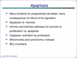
APOPTOSIS
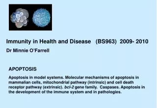
Immunity in Health and Disease (BS963) 2009- 2010 Dr Minnie O’Farrell . APOPTOSIS.

APOPTOSIS
E N D
Presentation Transcript
Immunity in Health and Disease (BS963) 2009- 2010 Dr Minnie O’Farrell APOPTOSIS Apoptosis in model systems. Molecular mechanisms of apoptosis in mammalian cells, mitochondrial pathway (intrinsic) and cell death receptor pathway (extrinsic). bcl-2 gene family. Caspases. Apoptosis in the development of the immune system and in pathologies.
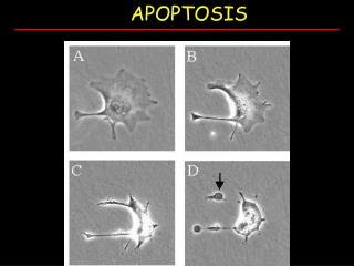
General text books/monographs: Alberts et al. The Molecular Biology of the Cell 5th ed Lodish et al. Molecular Cell Biology 6th ed. Weinberg The Biology of Cancer Normal white blood cell Apoptotic white blood cell Lodish 6th Fig 1-19 During apoptosis (programmed cell death) cells bleb and eventually break apart without releasing contents.
Alberts et al. Fig 18-3 Apoptosis during the metamorphosis of a tadpole into a frog. The cells in the tadpole tail are induced to undergo apoptosis stimulated by the increases in thyroid hormone that occurs during metamorphosis The nematode Caenorhabditis elegans has also been a very important model system for studying apoptosis in development.
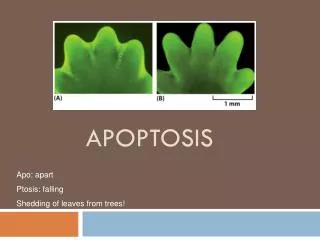
Types of cells that undergo apoptosis Weinberg Fig 9.19 Apoptosis and normal morphogenesis. TUNEL assay (immunodetection) detects breaks in DNA as cells undergo apoptosis Apoptosis in the embryonic mouse paw. Apoptosis occurs between 12.5 to 14.5 days in embryogenesis Cell Biology Pollard and Earnshaw + 1 day
Apoptosis is also important in the development of the nervous system
Two lymphocytes Staurosporin-treated HeLa cells Healthy Apoptotic Apoptosis Apoptosis Weinberg Fig 9.18 Different parts of the apoptotic programme. Markers for the process. Fragmentation of DNA Apoptotic cell Pyknotic nuclear fragments Phagocytosis of apoptotic bodies Fragmentation of Golgi bodies Phosphorylation of Histone 2B
Comparison between two forms of cell death, apoptosis and necrosis Apoptosis Necrosis Apoptosis is a highly regulated process requiring gene expression. Weinberg Table 9.3 Apoptosis v necrosis
Apoptosis is a very “clean” process. Phagocytic macrophage removing remains of apoptotic cell. Also a highly regulated process involving cell-cell interaction and signalling. Cell and Molecular Biology Karp
An important model system in which the molecular basis of apoptosis has been studied Lodish et al. 6th Fig 1.25
Caenorhabditis elegans Nomarski interference microscopy Lodish et al. 5th Fig 22.8 Lineage of all somatic cells, from fertilized egg to mature worm has been traced. 1030 cells generated but 131 cells die C. elegans Cell fate data
Genetic screens: Ced-3, Ced-4 and Egl1 required for cell deaths Ced-9 represses cell death programme ced-1 mutant (defective in engulfment so dead cells visible (refractile) and can identify apoptosed cells (yellow arrows) ced-1- ced-3 double mutant. There are no refractile cells in these double mutants indicating that no cell deaths occurred (yellow arrowheads) Mutations in the ced-3 gene block apoptosis Lodish et al.6th Fig 21.36
Lodish et al. 6th Figure 21.37 Evolutionary conservation of apoptotic pathways
There are two major pathways of apoptosis intrinsic pathway extrinsic pathway Maniati et al. 2008 Fig 1 The molecular basis of apoptosis in mammals. Simplified overview
A family of apoptosis proteins has been discovered in mammalian cells . The first member of the Bcl-2 family was identified during a study of B cell lymphoma. The oncogenic version is formed through a reciprocal chromosomal translocation in which parts of the chromosome 14 and chromosome 18 are exchanged. The translocated bcl-2 gene is now under the control of an active immunoglobulin promoter that drives high levels of constitutive expression. Bcl-2 is pro-survival (anti-apoptotic) and is homologous to CED-9 in Caenorhabditis elegans. Quite early in the study of Bcl-2 it was found to localise to the outer membrane of the mitochondria. We now know that there are at least 24 Bcl-2-related proteins, 6 are anti-apoptotic and 18 are pro-apoptotic. Lets examine these in a little more detail.
Introducing the BCL-2 family of proteins important in apoptotic pathways = anti-apoptotic Weinberg Fig 9.25 Bcl-2 and related proteins part A
The structure of many of these proteins has been determined and interactions between them investigated. Weinberg Fig 9.25 Bcl-2 and related proteins The anti-apoptotic protein, BCL-XL , is inhibited by binding of the pro-apoptotic BH3 only protein (orange) in the groove between BH1 and BH3
BH3 only protein binding specificity for BCL-2 homologues BIM and PUMA bind to all BCL-2 family members tested; by contrast NOXA only binds to A1 and MCL1. These binding specificities recapitulate the ability of these proteins to activate apoptosis e.g. BIM et al can induce apoptosis alone whereas a combination of NOXA and BAD is required. Youle and Strasser (2008) The BCL-2 protein family: opposing activities that mediate cell death. Nature Reviews Molecular Cell Biology, 9, 47-59
Conformational changes in BCL-2 family members during apoptosis. BAX undergoes extensive conformational changes during the mitochondrial translocation process. The protein changes from a soluble cytoplasmic protein in healthy cells to one that appears to have at least 3 helices inserted in the mitochondrial membrane in apoptotic cells. Youle and Strasser (2008) The BCL-2 protein family: opposing activities that mediate cell death. Nature Reviews Molecular Cell Biology, 9, 47-59
Alberts et al. Fig 18-7 Release of cytochrome c from mitochondria during apoptosis GFP = Green Fluorescent protein Control UV-treated to induce apoptosis
To summarize….. BCL-2 family of proteins have opposing apoptotic activities 1st set (e.g. Bcl-2 itself) inhibit apoptosis. 2nd set (e.g. BAX) promotes apoptosis. 3rd set (e.g. the BH3 only proteins) bind and regulate the anti-apoptotic BCL-2 proteins to promote apoptosis.
Extrinsic or death receptor pathway Maniati et al. 2008 Fig 1 The molecular basis of apoptosis. Simplified overview
Death receptors Weinberg Fig 9.31 The extrinsic apoptotic pathway
Maniati et al. 2008 Fig 1 The molecular basis of apoptosis. Simplified overview
Heptamer ( 7 chains) The blue helices in the middle bind and activate procaspase 9 to caspase 9 Caspase 9 activates procaspases 3, 6 and 7 Weinberg Fig 9.28 The APOPTOSOME is assembled in the cytoplasm when cytochrome c is released from the mitochondria and binds to Apaf-1
Weinberg Fig 9.32 Convergence of intrinsic and extrinsic apoptotic pathways
Caspases are proteolytic enzymes activated by both extrinsic and intrinsic pathways
Caspase family, 12-13 members Two classes: Initiators Effectors All caspases have a similar domain structure Not all mammalian caspases participate in apoptosis. For example Caspases 1, 4, 5, and 12 are activated during innate immune responses and are involved in the regulation of the inflammatory reponse
Effectorcaspases(such as caspase-3, -6 and -7 in mammals) function to breakdown cell structures through cleavage of specific substrates. Actin cytoskeleton Lamins Golgi Translation apparatus Taylor et al. (2008)
Phagocytic macrophage removing remains of apoptotic cell. This is a highly regulated process involving recognition and signalling between the apoptotic cell and macrophage. Cell and Molecular Biology Karp
Stages of engulfment of apoptotic cells can be divided into 4 stages Binding Recognition Phagocytosis Internalization Kinchen and Ravichandran (2007) Fig1
Lauber et al.(2004) Fig 2 Lack of “don’t eat me” signals on the surface of apoptotic cells
Lauber et al. (2004) Fig 6 The three steps of apoptotic cell removal.
Alberts et al. Fig 25-13 Mechanisms of immunological tolerance to self antigens
Alberts Fig 25-47 Two strategies by which effector cytotoxic T cells kill their target cells
Following are three supplementary slides for overview and glossary of terms. A useful diagramatic overview with a short review. Willis et al. (2003) J. Cell Science, 116, 4053-4057