PET imaging
This article explores the significance of positron emission tomography (PET) imaging, particularly the use of radioactive glucose as a radiotracer. It examines the process of creating radiotracers and how they interact within a magnetic field, emphasizing the principles of beta-plus and beta-minus decay. The paper details the stable and unstable isotopes involved, including F-18, and their applications in medical diagnostics. Adapted from the work of Jens Langner, this study offers a comprehensive look at the technology and science underpinning PET imaging.

PET imaging
E N D
Presentation Transcript
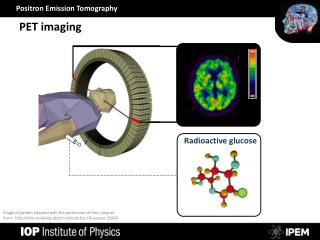
PET imaging Radioactive glucose Image of patient adapted with the permission of Jens Langner from: http://nbn-resolving.de/urn:nbn:de:bsz:14-qucosa-23509
Making radiotracers Radioactive glucose Electromagnet South pole Magnetic field O F 18 18 8 9 North pole Proton beam Target
Positron emmision beta-minus decay 11 Stable isotopes Unstable Stable 10 n p 9 Neutron Number beta-plus decay b + 8 18 17 16 O O O N F 16 18 positron (anti-particle of an electron) 8 8 8 9 7 7 6 6 7 8 9 10 Proton Number
Gamma pairs Gamma ray electron positron Gamma ray