Download
1 / 1
10 likes | 34 Views
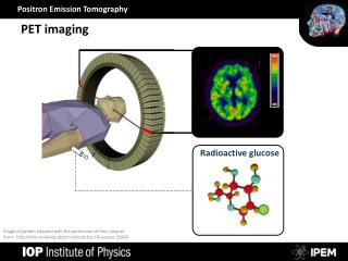
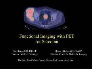
Atherosclerosis has become a major cause of cardiovascular diseases, mortality and morbidity. And the decisive factor lead to thrombosis is proved to be the rupture of vulnerable plaques. PET imaging are able to provide more accurate diagnosing values for atherosclerotic plaques, based on various pathophysiology /biomarkers. Nowadays, we had a increasing understanding of the utilization of different clinical radioprobes including 18F-FDG indicating for glucose metabolism in macrophage, 68Ga-DOTATATE binding somatostatin receptor on macrophage, 68Ga-Pentixafor affinity to C-X-C chemokine receptor type 4 (CXCR-4) in immune cells and 18F-NaF binding active arterial calcification; which are related to the different pathological process of atherosclerosis.

E N D
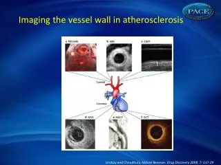
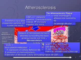
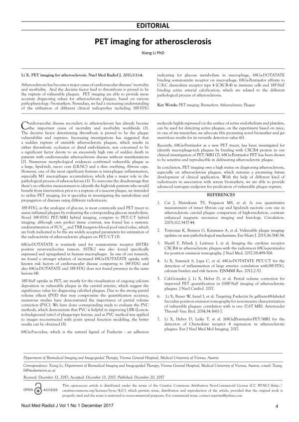
EDITORIAL PET imaging for atherosclerosis Xiang Li PhD Li X. PET imaging for atherosclerosis. Nucl Med Radiol J. 2017;1(1):4. indicating for glucose metabolism in macrophage, 68Ga-DOTATATE binding somatostatin receptor on macrophage, 68Ga-Pentixafor affinity to C-X-C chemokine receptor type 4 (CXCR-4) in immune cells and 18F-NaF binding active arterial calcification; which are related to the different pathological process of atherosclerosis. Atherosclerosis has become a major cause of cardiovascular diseases’ mortality and morbidity. And the decisive factor lead to thrombosis is proved to be the rupture of vulnerable plaques. PET imaging are able to provide more accurate diagnosing values for atherosclerotic plaques, based on various pathophysiology /biomarkers. Nowadays, we had a increasing understanding of the utilization of different clinical radioprobes including 18F-FDG Key Words: PET imaging; Biomarkers; Atherosclerosis; Plaques C The decisive factor determining thrombosis is proved to be the plaque vulnerability and ruptures. Increasing investigations has suggested that a sudden rupture of unstable atherosclerotic plaques, which results in either thrombotic occlusion or distal embolization, was concerned to be a significant factor devote to an excessively high rate of sudden death in patients with cardiovascular atherosclerosis disease without manifestations (2). Numerous morphological evidences confirmed vulnerable plaque as a large, lipid-rich, necro core (LR-NC) and a thin overlying, fibrous caps. However, one of the most significant features is intra-plaque inflammation, especially M1 macrophages accumulation; which play a major role in the pathological process of atherosclerosis (1). To overcome the disadvantage that there’s no effective measurement to identify the high-risk patients who would benefit from intervention prior to a rupture of a nascent plaque, we intended to utilize PET imaging, for it specialize in investigating the metabolism and propagation of diseases using different radiotracers. molecule highly expressed on the surface of active endothelium and platelets, can be used for detecting active plaques, on the experiment based on mice, in one of my researches, we advocate this promising novel biomarker and get marvelous results for its versatile detection value (6). ardiovascular disease secondary to atherosclerosis has already became the important cause of mortality and morbidity worldwide (1). Recently, 68Ga-Pentixafor as a new PET tracer, has been investigated for identify macrophage-rich plaques by binding with CXCR4 protein in our clinical investigation of PET/MRI (7). 68Ga-Pentixafor PET has been shown to be sensitive and reproducible in delineating atherosclerotic plaque. In conclusion, PET imaging own a high status on diagnosing atherosclerosis, especially on atherosclerosis plaques, which remains a promising future development of clinical application. With the help of different kind of radiotracers in association with serum biomarkers, we are able to provide advanced surrogate endpoint for predication of vulnerable plaque rupture. REFERENCES 1. Cai J, Hatsukami TS, Ferguson MS, et al. In vivo quantitative measurement of intact fibrous cap and lipid-rich necrotic core size in atherosclerotic carotid plaque: comparison of high-resolution, contrast- enhanced magnetic resonance imaging and histology. Circulation. 2005;112:3437-44. 18F-FDG, as the analogue of glucose, is most commonly used PET tracer to assess inflamed plaques by evaluating the corresponding glucose metabolism. Novel 18F-FDG PET/MRI hybrid imaging, compare to PET/CT hybrid imaging, although own perfect tissue contrast, was found has a systemic underestimation of SUVmax and TBR (target-to-blood pool ratio) value, which are both indicated to be the are widely accepted parameters for estimation of the radioactivity of atherosclerotic plaques on PET/CT (3). 2. Toutouzas K, Benetos G, Karanasos A, et al. Vulnerable plaque imaging: updates on new pathobiological mechanisms. Eur Heart J. 2015;36:3147-54. 3. Hyafil F, Pelisek J, Laitinen I, et al. Imaging the cytokine receptor CXCR4 in atherosclerotic plaques with the radiotracer 68Ga-pentixafor for positron emission tomography. J Nucl Med. 2017;58:499-506 68Ga-DOTATATE is routinely used for somatostatin receptor (SSTR)- positive neuroendocrine tumors. SSTR-2 was also found specifically expressed and upregulated in human macrophages. In one of our research, we found a stronger relation of increased 68Ga-DOTATATE uptake with known risk factors of cardiovascular disease comparing to 18F-FDG and also 68Ga-DOTATATE and 18F-FDG does not found presence in the same lesions (4). 4. Li X, Samnick S, Lapa C, et al. 68Ga-DOTATATE PET/CT for the detection of inflammation of large arteries: correlation with18F-FDG, calcium burden and risk factors. EJNMMI Res. 2012;2:52. 5. Cal-Gonzalez J, Li X, Heber D, et al. Partial volume correction for improved PET quantification in (18)F-NaF imaging of atherosclerotic plaques. J Nucl Cardiol. 2017. 18F-NaF uptake in PET, are mostly for the visualization of ongoing calcium deposition in vulnerable plaque in the carotid arteries, which suggest the significance value for diagnosing calcified plaques. Due to the strong partial volume effects (PVE) that may compromise the quantification accuracy, numerous studies have demonstrated the importance of partial volume correction (PVC). We have done corresponding study to evaluate the PVC methods, which demonstrate that PVC is helpful in improving LBR (Lesion- to-background ratio) of plaque-type lesions, and as PVC method was applied to images reconstructed with point spread function modeling, the better results can be obtained (5). 6. Li X, Bauer W, Israel I, et al. Targeting P-selectin by gallium-68-labeled fucoidan positron emission tomography for non-invasive characterization of vulnerable plaques: correlation with in vivo 17.6T MRI. Arterioscler Thromb Vasc Biol. 2014;34:1661-7. 7. Li X, Heber D, Leike T, et al. [68Ga]Pentixafor-PET/MRI for the detection of Chemokine receptor 4 expression in atherosclerotic plaques. Eur J Nucl Med Mol Imaging. 2017. 68Ga-Fucoidan, which is the natural ligand of P-selectin - an adhesion Department of Biomedical Imaging and Image-guided Therapy, Vienna General Hospital, Medical University of Vienna, Austria Correspondence: Xiang Li, Department of Biomedical Imaging and Image-guided Therapy, Vienna General Hospital, Medical University of Vienna, Austria. e-mail: Xiang. li@meduniwien.ac.at Received: December 12, 2017; Accepted: December 13, 2017; Published: December 20, 2017 This open-access article is distributed under the terms of the Creative Commons Attribution Non-Commercial License (CC BY-NC) (http:// creativecommons.org/licenses/by-nc/4.0/), which permits reuse, distribution and reproduction of the article, provided that the original work is properly cited and the reuse is restricted to noncommercial purposes. For commercial reuse, contact reprints@pulsus.com OPEN ACCESS Nucl Med Radiol J Vol 1 No 1 December 2017 4