Download
1 / 18
180 likes | 283 Views
This study presents a novel methodology for reconstructing 3D dense cardiac motion from tagged MRI sequences. By integrating prior knowledge of myocardial fiber structure with advanced continuum mechanics, we apply constrained energy minimization to enhance the accuracy of cardiac motion modeling. The reconstruction process utilizes a fiber-based model to establish correspondences between transverse MRI slices, allowing for detailed analysis of myocardial deformation in both healthy and pathological states. Results demonstrate significant advancements in the understanding of cardiac mechanics, with promising applications in cardiovascular research.

E N D
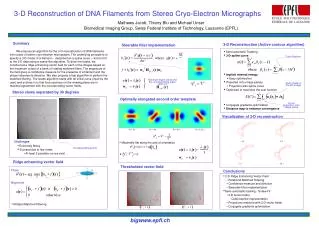
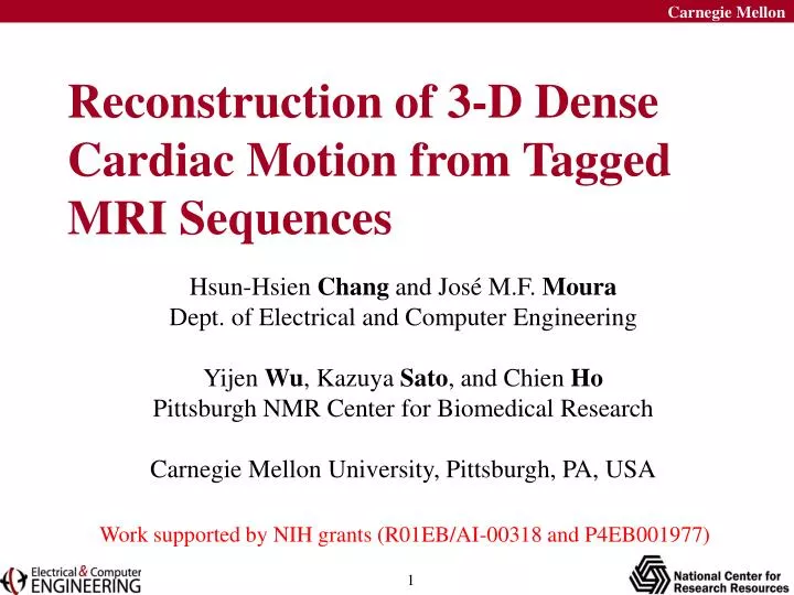
Reconstruction of 3-D Dense Cardiac Motion from Tagged MRI Sequences Hsun-Hsien Chang and José M.F. Moura Dept. of Electrical and Computer Engineering Yijen Wu, Kazuya Sato, and Chien Ho Pittsburgh NMR Center for Biomedical Research Carnegie Mellon University, Pittsburgh, PA, USA Work supported by NIH grants (R01EB/AI-00318 and P4EB001977)
Outline • Introduction • Methodology: Prior knowledge + MRI data • Myocardial Fiber Based Structure • Continuum Mechanics • Constrained Energy Minimization • Results and Conclusions
N slices 2-D Cardiac MRI Images M frames per slice sparse displacements dense displacements Y. Sun, Y.L. Wu, K. Sato, C. Ho, and J.M.F. Moura, Proc. Annual Meeting ISMRM 2003
3-D Reconstruction: myocardial fiber model Use a fiber based model to find the correspondence between transversal slices.
Use continuum mechanics to describe the motion of fibers. Fit the model to MRI data by constrained energy minimization 3-D Reconstruction: fiber deformation model
Outline • Introduction • Methodology: Prior knowledge + MRI data • Myocardial Fiber Based Structure • Continuum Mechanics • Constrained Energy Minimization • Results and Conclusions
+60º -60º Prior Knowledge: myocardial anatomy Endocardium Multiple-layer view: Mid-wall Epicardium Streeter, in Handbook of Physiology Volume 1: the Cardiovascular System, American Physiological Society, 1979
a(t)+da(t) da(t) a(t) Displacement: u(t)=a(t)-a(0) Prior Knowledge: fiber dynamics Motion of a small segment Notations are column vectors, ex: da(0) a(0)+da(0) a(0)
Deformation Gradient Matrix Deformation gradient F(t) is a function of displacement u(t).
Strain • Strain is the displacement per unit length, and is written mathematically as Ref: Y.C. Fung, A First Course in Continuum Mechanics, 3rd ed., Prentice-Hall, New Jersey, 1994 • When strain is small, it is approximated as (Note: S is symmetric)
Linear Strain Energy Model • S is symmetric, so we vectorize the entries at upper triangle. • Let C describe the material properties. It can be shown the linear strain energy is • The entire energy of the heart:
Constrained Energy Minimization • Internal energy: continuum mechanics governs the fibers to move as smooth as possible. • External energy: pixel intensities of fibers should be kept similar across time.
2-D Displacement Constraints D: 2-D displacements of the taglines ӨU: picks the entries of U corresponding to D 2-D displacement constraints: ӨU=D λ: Lagrange multiplier
Outline • Introduction • Methodology: Prior knowledge + MRI data • Myocardial Fiber Based Structure • Continuum Mechanics • Constrained Energy Minimization • Results and Conclusions
10 frames per slice Data Set 4 slices 256256 pixels per image • Transplanted rats with heterotropic working hearts. • MRI scans performed on a Bruker AVANCE DRX 4.7-T system Y. Sun, Y.L. Wu, K. Sato, C. Ho, and J.M.F. Moura, Proc. Annual Meeting ISMRM 2003
Fiber Based Model Whole left ventricle epicardium endocardium mid-wall
Conclusions • Take into account the myocardial fiber based structure. • Adopt the continuum mechanics framework. • Implement constrained energy minimization algorithms.