Download

1 / 59
670 likes | 1.38k Views
CARDIAC MRI. Diagnostic Backgrounder. NOTE: These slides are for use in educational oral presentations only. If any published figures/tables from these slides are

E N D
CARDIAC MRI Diagnostic Backgrounder NOTE: These slides are for use in educational oral presentations only. If any published figures/tables from these slides are to be used for another purpose (e.g. in printed materials), it is the individual’s responsibility to apply for the relevant permission.Specific local use requires local approval
Outline • Introduction to iron overload • Assessing cardiac iron loading • echocardiography • cardiac MRI • Cardiac MRI in practice • preparation of the patient • acquisition of the image • analysis of the data • Excel spreadsheet • ThalassaemiaTools (CMRtools) • cmr42 • FerriScan • MRmap • MATLAB • Summary MRI = magnetic resonance imaging.
Introduction to iron overload • Iron overload is common in patients who require intermittent or regular blood transfusions to treat anaemia and associated conditions • it may be exacerbated in some conditions by excess gastrointestinal absorption of iron • Iron overload can lead to considerable morbidity and mortality1 • Excess iron is deposited in major organs, resulting in organ damage • the organs that are at risk of damage due to iron overload include the liver, heart, pancreas, thyroid, pituitary gland, and other endocrine organs2,3 1Ladis V, et al. Ann NY Acad Sci. 2005;1054:445-50. 2Gabutti V, Piga A. ActaHaematol. 1996;95:26-36. 3Olivieri NF. N Engl J Med. 1999;341:99-109.
Importance of analysing cardiac iron • In β-thalassaemia major, cardiac failure and arrhythmia are risk factors for mortality1 • signs of myocardial damage due to iron overload: arrhythmia, cardiomegaly, heart failure, and pericarditis2 • heart failure has been a major cause of death in β-thalassaemia patients in the past (50–70%)1,3 • In MDS, the results of studies are less comprehensible • the reported proportion of MDS patients with cardiac iron overload is inconsistent; from high to only a small proportion of MDS patients4–7 • cardiac iron overload occurs later than does liver iron overload4,7,8 • however, cardiac iron overload can have serious clinical consequences in MDS patients 1Borgna-Pignatti C, et al. Haematologica. 2004;89:1187-93. 2Gabutti V, Piga A. ActaHaematol. 1996;95:26-36. 3. Modell B, et al. Lancet. 2000;355:2051-2. 4Jensen PD, et al. Blood. 2003;101:4632-9. 5Chacko J, et al. Br J Haematol. 2007;138:587-93. 6Konen E, et al. Am J Hematol. 2007;82:1013-6. 7Di Tucci AA, et al. Haematologica. 2008;93:1385-8. 8Buja LM, Roberts WC. Am J Med. 1971;51:209-21.
70 60 60 50 40 30 23 17 20 10 7 0 Importance of analysing cardiac iron (cont.) • In 2010, the overall mortality rate of β-thalassaemia major patients in the UK was substantially lower than a decade ago (1.65 vs 4.3 per 1,000 patient years)1,2 • due to improved monitoring and management of iron overload over the last decade, 77% of patients have normal cardiac T2*1 • cardiac iron overload is no longer the leading cause of death in this population1 Baseline Latest follow-up Patients (%) p < 0.001 p < 0.001 cT2* < 10 ms cT2* ≤ 20 ms cT2* = cardiac T2*. 1Thomas AS, et al. Blood. 2010;116:[abstract 1011]. 2Modell B, et al. Lancet. 2000;355:2051-2.
Cardiac T2*: Overview of correlations with other measurements Transfusion duration†↑1 Ventricular dysfunction↑1-3 Arrhythmia and heart failure↑4 T2*↓ Need for cardiac medication↑1-2 APFR↓EPFR:APFR↑5 SF and LIC1-3 Weak or no correlation †For thalassaemia, but not sickle cell. APFR = atrial peak filling rate; EPFR = early peak filling rate; LIC = liver iron concentration; SF = serum ferritin. 1Wood JC, et al. Blood. 2004;103:1934-6. 2Anderson LJ, et al. Eur Heart J. 2001;22:2171-9. 3Tanner MA, et al. J CardiovascMagnReson. 2006;8:543-7. 4Kirk P, et al. Circulation. 2009;120:1961-8. 5Westwood MA, et al. J MagnReson Imaging. 2005;22:229-33.
Cardiac T2*: Relationship with LVEF Normal T2* range 90 80 Normal LVEF range 70 60 Cardiac T2* value of 37 ms in a normal heart 50 LVEF (%) 40 30 20 10 0 Cardiac T2* value of 4 ms in a significantly iron-overloaded heart 0 10 20 30 40 50 60 70 80 90 100 Cardiac T2* (ms) Myocardial T2* values < 20 ms are associated with a progressive and significant decline in LVEF LVEF = left-ventricular ejection fraction. Anderson LJ, et al. Eur Heart J. 2001;22:2171-9.
0.30 0.25 0.20 0.15 0.10 0.05 0 Cardiac T2*: Relationship with cardiac failure and arrhythmia Cardiac failure Arrhythmia 0.6 < 6 ms 0.5 < 10 ms 0.4 6–8 ms Proportion of patients with arrhythmia Proportion of patients developing cardiac failure 0.3 0.2 8–10 ms 10–20 ms 0.1 > 20 ms > 10 ms 0 0 60 120 180 240 300 360 0 60 120 180 240 300 360 Follow-up time (days) Follow-up time (days) T2* < 10 ms: relative risk 159 (p < 0.001)T2* < 6 ms: relative risk 268 (p < 0.001) T2* < 20 ms: relative risk 4.6 (p < 0.001)T2* < 6 ms: relative risk 8.65 (p < 0.001) Low myocardial T2* predicts a high risk of developing cardiac failure and arrhythmia Kirk P, et al. Circulation. 2009;120:1961-8.
Assessing cardiac iron loading: Agenda • Echocardiography • Cardiac MRI • advantages and disadvantages of cardiac MRI • MRI: a non-invasive diagnostic tool • T2* is the standard method for analysing cardiac iron
Assessing cardiac iron loading: Echocardiography EF = ejection fraction. 1Leonardi B, et al. JACC Cardiovasc Imaging. 2008;1:572-8. 2Hoffbrand AV. Eur Heart J. 2001;22:2140-1.
MRI: A non-invasive diagnostic tool Protons • Indirectly measures levels of iron in the heart • MRI measures longitudinal (T1) and transverse (T2) relaxation times of the protons • iron deposition disrupts the homogeneous magnetic field and shortens T1 and T2 times in a concentration-dependent manner Magnetic field RF/spin echo/gradient echo Iron Echo signal → T1, T2 Signal processing RF = radio-frequency. 1Wood JC, Ghugre N. Hemoglobin. 2008;32:85-96. 2Wood JC, et al. Circulation. 2005;112:535-43. 3Wang ZJ, et al. Radiology. 2005;234:749-55. 4Ghugre NR, et al. MagnReson Med. 2006;56:681-6.
MRI: A non-invasive diagnostic tool (cont.) Protons • If a spin-echo sequence is used, the relaxation time is T2 • If a gradient-echo sequence is used, it is T2* • Cardiac MRI methods • gradient-echo T2* MRI: most used in clinical practice • spin-echo T2 MRI: less useful (motion artefacts common due to characteristics of the heart) Magnetic field Most used in clinical practice: Gradient echo Spin echo Image acquired at different TEs Image acquired at different TEs Excel or software Excel or software T2* [ms} T2* [ms} R2* [Hz]=1,000/T2* R2* [Hz]=1,000/T2* TE = echo time. Adapted from Wood JC, Ghugre N. Hemoglobin. 2008;32:85-96.
FAQ: Cardiac MRI What are sequences? Sequences are a set of radio-frequency and gradient pulses (slight tilts in the magnetization curves of the scanner) generated repeatedly during the scan, which produce echoes with varied amplitudes and shapes that will define the MR image What is gradient echo? A gradient-echo sequence is obtained after 2 gradient impulses are applied to the body, resulting in a signal echo that is read by the coils. In these sequences, the spins are not refocused and, therefore, are subject to local inhomogeneities, with a more rapid decay curve. For gradient-echo pulse sequences, the T2* relaxation times (which reflect these inhomogeneities) on the signal are more significant 1Image from Ridgway JP. J CardiovascMagnReson. 2010;12:71.
Gradient relaxometry (T2*, R2*) is the method for analysing cardiac iron levels 1Guo H, et al. J MagnReson Imaging. 2009;30:394-400. 2Anderson LJ, et al. Eur Heart J. 2001;22:2171-9. 3Wood JC, Noetzli L. Ann N Y Acad Sci. 2010;1202:173-9.4Wood JC, Ghugre N. Hemoglobin. 2008;32:85-96. 5Westwood M, et al. J MagnReson Imaging. 2003;18:33-9. 6Hoffbrand AV. Eur Heart J. 2001;22:2140-1. 7He T, et al. MagnReson Med. 2008;60:1082-9.
14 12 10 8 6 4 2 0 0 100 200 300 400 Gradient relaxometry (T2*, R2*) can conveniently measure cardiac and liver iron Liver MRI Cardiac MRI 30 Hankins, et al. 25 20 Wood, et al. 15 HIC (mg Fe/g of dry weight liver) [Fe] (mg/g dry wt) 10 Anderson, et al. R2 = 0.82540 5 0 0 200 400 600 800 1000 Cardiac R2* (Hz) Liver R2* (Hz) Cardiac and liver iron can be assessed together conveniently by gradient echo during the a single MRI measurement. HIC = hepatic iron concentration Carpenter JP, et al. J CardiovascMagnReson. 2009;11 Suppl 1:P224. Hankins et al Blood. 2009;113:4853-4855.
Cardiac T2* MRI is usually measured in the septum of the heart Heart with normal iron levels T2* = 22.8 ms or R2* = 43.9 Hz Heart with severe iron overload T2* = 5.2 ms or R2* = 192 Hz Images courtesy of Dr J. de Lara Fernandes.
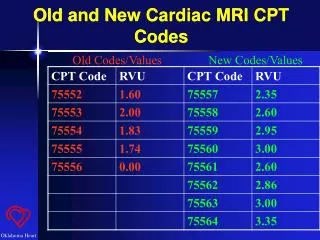
What is R2*? Conversion from T2* to R2* is a simple mathematical calculation: R2* = 1,000/T2* These values are only applicable to 1.5 T scanners1 1Anderson LJ, et al. Eur Heart J. 2001;22:2171-9. 2Kirk P, et al. Circulation. 2009;120:1961-8.
14 12 10 8 6 14 12 4 10 2 8 6 0 4 0 10 20 30 40 50 60 70 2 0 0 100 200 300 400 Why should the data be presented as R2* and not T2*? • Seven whole hearts from patients with transfusion-dependent anaemias were assessed by histology and cardiac MRI [Fe] (mg/g dry wt) [Fe] (mg/g dry wt) R2 = 0.949 R2 = 0.82540 Cardiac T2* (ms) Cardiac R2* (Hz) R2* has a linear relationship with tissue iron concentration, which simplifies the interpretation of data and allows comparison of changes over time Carpenter JP, et al. J CardiovascMagnReson. 2009;11 Suppl 1:P224.
100 80 60 40 20 0 Why should the data be presented as R2* and not T2*? (cont.) The relationship between cardiac T2*/R2* and LVEF Hockey stick effect? Or a more gradual relationship? 90 80 70 60 50 LVEF (%) LVEF (%) 40 30 20 10 0 0 50 100 150 200 250 10 20 30 40 50 60 70 80 90 100 0 Heart T2* (ms) R2* (s–1) R2* allows demonstration of cardiac risk in a more gradual way Anderson LJ, et al. Eur Heart J. 2001;22:2171-9.
Why should the data be presented as R2* and not T2*? (cont.) Standard errors on a single measurement are approximately constant with R2*, but are non-uniform with T2* Transform to R2* 60 120 50 100 40 80 T2* first measurement (ms) R2* first measurement (s–1) 30 60 20 40 20 10 0 0 0 10 20 30 40 50 60 0 20 40 60 80 100 120 T2* second measurement (ms) R2* second measurement (s–1) R2* has a constant standard error that makes assessment of the significance of changes easier Westwood M, et al. J MagnReson Imaging. 2003;18:33-9.
MRI scanners • Manufacturers • Siemens Healthcare (Erlangen, Germany; www.siemensmedical.com) • GE Healthcare (Milwaukee, WI, USA; www.gemedicalsystems.com) • Philips Healthcare (Best, the Netherlands; www.medical.philips.com) • Magnetic field • T2* varies with magnetic field strength1 • need 1.5 T for cutoff levels of 20 ms (iron overload) and 10 ms (severe iron overload)1,2 • Cardiac package • needs to be acquired separately from the manufacturers. The cost is about USD 40,000. However, in most centres, this is available since MRI is frequently used in non-iron-related cardiovascular imaging • includes all necessary for acquisition of the image • sequences are included in Siemens and Philips Healthcare cardiac packages, but for GE Healthcare they need to be acquired separately (note: variations may exist between countries) 1Anderson LJ, et al. Eur Heart J. 2001;22:2171-9. 2Kirk P, et al. Circulation. 2009;120:1961-8.
Cardiac T2* MRI in practice: The process 3. Analysis ofMRI data(time depends on experience*) 2. Acquisition of the MRI image (approx. 5-20 min) 1. Patientpreparation(5 min) T2*, R2* *Time to manually calculate T2*/R2* values in an Excel spreadsheet depends on the experience of the physician.
Cardiac T2* MRI in practice: The process (cont.) • Preparation of the patient • Acquisition of the image • Analysis of the data (post-processing) • Excel spreadsheet • ThalassaemiaTools, CMRtools • cmr42 • FerriScan • MRmap • MATLAB
Preparation of the patient • Standard precautions need to be taken • There is no need for peripheral vein access since no contrast agent is required • Special care • remove all infusion/medication pumps (e.g. with insulin,pain-relieving drugs) • stop continuous i.v. application of ICT during the measurement • ECG signal should be positioned according to scanner specifications ECG = electrocardiography.
Cardiac T2* MRI in practice: The process (cont.) • Preparation of the patient • Acquisition of the image • Analysis of the data (post-processing) • Excel spreadsheet • ThalassaemiaTools, CMRtools • cmr42 • FerriScan • MRmap • MATLAB
Acquisition of the image: MRI pulse sequences • Pulse sequences • are a preselected set of defined radio-frequency and gradient pulses • are computer programs that control all hardware aspects of the scan • determine the order, spacing, and type of radio-frequency pulses that produce magnetic resonance images according to changes in the gradients of the magnetic field • Several different pulse sequences exist1 • a gradient-echo sequence generates T2* 1Wood JC, Ghugre N. Hemoglobin. 2008;32:85-96.
The most common commercially available T2* acquisition techniques The various techniques give clinically comparable results.2-3, 5 1Anderson LJ, et al. Eur Heart J. 2001;22:2171-9. 2Westwood M, et al. J Magn Reson Imaging. 2003;18:33-9. 3He T, et al. J Magn Reson Imaging. 2007;25:1205-9. 4He T, et al. Magn Reson Med. 2008;60:1082-9. 5Pepe A, et al. J Magn Reson Imaging. 2006;23:662-8.
Shortest TE = 2 ms Shortest TE = 1 ms 500 Shortest TE = 4 ms 450 Shortest TE = 5.5 ms True 400 350 Mean R2*: ramp, dualtone, & uniform (Hz) 300 250 200 150 100 50 0 100 200 300 400 500 0 True R2* (Hz) Acquisition of the image: TEs • Images are taken at a minimum of 5 different TEs, normally 8‒121 • The choice of minimum TE determines the smallest measurable T21 • ideally, min TE 2 ms, max TE 17‒20 ms • Different T2* acquisition techniques according to TE • multiple breath-hold: acquire an image for each TE in separate breath-holds2 • single breath-hold multi-echo acquisition: acquire images for all TE during 1 breath-hold3 Mean R2* compared with true value in the case of synthetic images for different minimum TEs, but same echo duration (18 ms)4 1Wood JC, Noetzli L. Ann N Y AcadSci. 2010;1202:173-9. 2Anderson LJ, et al. Eur Heart J. 2001;22:2171-9. 3Westwood M, et al. J MagnReson Imaging. 2003;18:33-9. 4Ghugre NR, et al. J MagnReson Imaging. 2006;23:9-16.
How does the MRI data output looks like? MRI data output Data visualization During a single breath hold the pulse sequence run several times at increasing echo time (TE), generating data points corresponding to decreased signal intensity1 1Wood JC, Ghugre N. Hemoglobin. 2008;32:85-96.
FAQ: Acquisition technique Which is recommended: single or multiple breath-hold technique? Comparison of the 2 methods, single and multiple breath-hold, showed no significant skewing between T2* values in all patients with -thalassaemia major, regardless of their T2* value (see Bland-Altman plots)1 However, in cardiac MRI the most recommended technique is single breath-hold, because it allows quick acquisition of the information. This is especially important to avoid movement artefacts (heart beating, breathing) and assure the good quality of the MRI image Patients with T2* < 20 ms1 Patients with T2* 20 ms 1 1Westwood M, et al. J MagnReson Imaging. 2003;18:33-9.
Acquisition of the image • Single breath-hold multi-echo acquisition • take a short-axis slice of the ventricle (halfway between the base and the apex): orange line • image acquisition should occur immediately after the R wave • do not alter any settings that could alter TE (e.g. FOV) Image courtesy of Dr J. de Lara Fernandes.
Cardiac T2* MRI in practice: The process (cont.) • Preparation of the patient • Acquisition of the image • Analysis of the data (post-processing) • Excel spreadsheet • ThalassaemiaTools, CMRtools • cmr42 • FerriScan • MRmap • MATLAB
How T2* is calculated from the MRI output? Data visualization Curve Fitting T2* Noise level T2* calculation is fitting a curve on the data points and calculating at what echo time no signal is left from iron (only noise)1 1Wood JC, Ghugre N. Hemoglobin. 2008;32:85-96.
Analysis of the data • The data can be analysed manually or using post-processing software
Analysis of the data (cont.) FDA = Food and Drug Administration. 1www.cmrtools.com/cmrweb/ThalassaemiaToolsIntroduction.htm. Accessed Dec 2010. 2Pennell DJ. JACC Cardiovasc Imaging. 2008;1:579-81.3www.circlecvi.com. Accessed Dec 2010.
Analysis of the data (cont.) 1www.resonancehealth.com/resonance/ferriscan. Accessed Dec 2010. 2www.cmr-berlin.org/forschung/ mrmapengl/index.html. Accessed Dec 2010. 3Wood JC, Noetzli L. Ann N Y Acad Sci. 2010;1202:173-9.
FAQ: Mistakes in analysing the data What are the most common mistakes in analysing the data that could lead to a wrong interpretation of the T2* value? • Interpreting the data from cardiac MRI is usually quite straightforward; problems may arise when analysing data from patients with severe cardiac iron overload. In this case, the signal from heavily iron-loaded muscle will decay quickly and a single exponential decay curve does not fit the data well.1 • Models exist that can help to solve this issue (see next slide): • the offset model (Prof Wood and colleagues) • truncation of the data (Prof Pennell and colleagues) • Both models should give comparable results; the differences should not be clinically relevant Signal decay curve from a patient with T2* ≈ 5 ms, showing that the data do not fit well2 1Wood JC, Noetzli L. Ann N Y Acad Sci. 2010;1202:173-9. 2Ghugre NR, et al. J MagnReson Imaging. 2006;23:9-16.
FAQ: Mistakes in analysing the data (cont.) What is truncation? After the selection of the ROI, the signal decay can be fitted using different models. In the truncation model, the late points in the curve that form a plateau are subjectively discarded; the objective is to have a curve with an R2 > 0.995. A new single exponential curve is made by fitting the remaining signals.1 Generally, a truncation model should be used with the bright-blood technique to obtain more reproducible and more accurate T2* measurements1 What is an offset model? The offset model consists of a single exponential with a constant offset. Using only the exponential model can underestimate the real T2* values (at quick signal loss at short TE, there is a plateau), while inclusion of the offset model into the fitting equation can improve this.2 Generally, the offset model is recommended to be used with the black-blood technique 1He T, et al. MagnReson Med. 2008;60:1082-9. 2Ghugre NR, et al. J MagnReson Imaging. 2006;23:9-16.
FAQ: How to start measuring cardiac iron loading? How to start measuring cardiac iron loading in a hospital? What steps need to be taken? • To start assessing cardiac iron loading by MRI, these steps can be followed: • Check MRI machine requirements • 1.5 T • calibrated • Buy cardiac package from the manufacturer. It must include all that is necessary for acquisition of the data (the sequences are included with Siemens and Philips Healthcare cardiac packages, but for GE Healthcare they need to be acquired separately) • Optional: buy software for analysing the data (if not, Excel spreadsheet can be used) • Highly recommended: training of personnel for acquisition of cardiac MR images (e.g. functional analyses) • Highly recommended: training of personnel on how to analyse the data with the chosen software
Implementation of liver and cardiac MRI 1.5T MRI Scanner US$1.000.000 Yes ½ day training Liver Analysis Experienced radiologist No 1 day training Post-processing analysis US$40.000 or US$4.000/y or in-house or outsource US$50.000 Cardiac acquisition package 1-2 day training Yes Heart Analysis Routine cardiac MR exams 4 day training No Slide presented at Global Iron Summit 2011 - With the permission of Juliano de Lara Fernandes