Download

1 / 35
350 likes | 397 Views
Learn about the intricate development of the thyroid gland and tongue in the embryo, including histogenesis, thyroglossal duct cysts, and the formation of the posterior third of the tongue.

E N D
THYROID GLAND & TONGUE By: Dr. Mujahid Khan
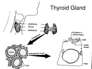
Development of Thyroid • The thyroid gland is the first endocrine gland to develop in embryo • It begins to form about 24 days after fertilization • It develops from a median endodermal thickening in the floor of a primordial pharynx • Thickening soon forms a small outpouching called thyroid primordium
Development of Thyroid • As the embryo and tongue grow, the developing thyroid gland descends in the neck, passing ventral to the developing hyoid bone and laryngeal cartilages • For a short time the thyroid gland is connected to the tongue by a narrow tube, the thyroglossal duct
Development of Thyroid • At first the thyroid primordium is hollow but it soon becomes solid and divides into right and left lobes • The two lobes are connected by the isthmus of the thyroid gland • Isthmus lies anterior to the developing second and third tracheal rings • By seventh week it assumes the definitive shape and has reached its final site in the neck
Development of Thyroid • The thyroglossal duct has normally degenerated by seventh week • The proximal opening of the thyroglossal duct persists as a small pit in the tongue, the foramen cecum • A pyramidal lobe extends upward from the isthmus in about 50% of people
Development of Thyroid • The pyramidal lobe may be attached to the hyoid bone by fibrous tissue or smooth muscle, the levator of thyroid gland • The pyramidal lobe and the associated smooth muscle represent a persistent part of the distal end of the thyroglossal duct
Histogenesis of Thyroid • The thyroid primordium consists of a solid mass of endodermal cells • The cellular aggregation later breaks up into a network of epithelial cords • By the tenth week the cords have divided into small cellular groups • A lumen soon forms in each cell cluster and the cells become arranged in a single layer around the lumen • During the eleventh week colloid begins to appear in these structures, called thyroid follicles • Iodine concentration and synthesis of thyroid hormones can be demonstrated
Thyroglossal Duct Cysts & Sinuses • Cyst may form anywhere along the course followed by the thyroglossal duct during descent of the primordial thyroid gland from the tongue • Normally the thyroglossal duct atrophies and disappear • A remnant of it may persist and form a cyst in the tongue or in the anterior part of the neck
Thyroglossal Duct Cysts & Sinuses • It usually lies just inferior to the hyoid bone • Most thyroglossal duct cysts are observed by the age of 5 years • The swelling produced is usually develops as a painless, progressively enlarging, moveable mass • The cyst may contain some thyroid tissue
Thyroglossal Duct Cysts & Sinuses • Following infection of a cyst, a perforation of the skin occurs forming a thyroglossal duct sinus • It usually opens in the median plane of the neck, anterior to the laryngeal cartilages
Development of Tongue • A median triangular elevation appears in the floor of the primordium pharynx near the end of 4th week, just rostral to the foramen cecum • This swelling or median tongue bud is the first indication of tongue development • Soon two oval distal tongue buds develop on each side of the median tongue bud
Development of Tongue • The three lingual buds result from the proliferation of mesenchyme in ventromedial parts of the first pair of pharyngeal arches • The distal tongue buds rapidly increase in size, merge with each other, and overgrow the median tongue bud • The merged distal tongue buds form the anterior two-thirds (oral part) of the tongue
Development of Tongue • Fusion of the distal tongue buds is indicated by a middle groove, the median sulcus of the tongue and internally by the fibrous lingual septum • Median tongue bud forms no recognizable part of the adult tongue
Formation of Posterior third of Tongue • It is indicated by two elevations that develop caudal to the foramen cecum • Copula: Forms by fusion of the ventromedial part of the second pair of pharyngeal arches • The hypopharyngeal eminence: develops caudal to the copula from mesenchyme in the ventromedial parts of the third and fourth pairs of arches
Formation of Posterior third of Tongue • As the tongue develops the copula is gradually overgrown by the hypopharyngeal eminence and disappear • As a result, the pharyngeal part of the tongue develops from the rostral part of the hypopharyngeal eminence • The line of fusion of the anterior and posterior parts of the tongue is roughly indicated by a V-shaped groove called terminal sulcus
Formation of Posterior third of Tongue • Pharyngeal mesenchyme forms the connective tissue and vasculature of the tongue • Most of the tongue muscles are derived from myoblasts that migrate from the occipital myotomes • The hypoglossal nerve (CN Ⅻ) accompanies the myoblast during their migration and innervates the tongue muscles as they develop • The entire tongue is within the mouth at birth, its posterior third descends into the oropharynx by 4 years of age
Papillae and Taste Buds • Lingual papillae appear towards the end of the eighth week • The vallate and foliate papillae appear first, close to the terminal branches of the glossopharyngeal nerve (CN Ⅸ) • The fungiform papillae appear later near termination of chorda tympani branch of the facial nerve
Papillae and Taste Buds • The most common lingual papillae, known as filiform papillae because of their threadlike shape, develop during early fetal period (10-11 weeks) • They contain afferent nerve endings sensitive to touch • Taste buds develop during 11-13 weeks • Most taste buds form on the dorsal surface of the tongue
Papillae and Taste Buds • Fetal responses in the face can be induced by bitter tasting substances at 26-28 weeks, indicating that the reflex pathways between taste buds and facial muscles are established by this age
Nerve Supply of the Tongue • The development of tongue explains its nerve supply • The sensory supply to the mucosa of almost the entire anterior two-thirds of the tongue is from the lingual branch of the mandibular division of the trigeminal nerve • This nerve is the nerve of first pharyngeal arch and this arch forms the median and distal tongue buds
Nerve Supply of the Tongue • Facial nerve is the nerve of second pharyngeal arch • Its chorda tympani branch supplies the taste buds in the anterior two-thirds of the tongue except the vallate papillae • The facial nerve does not supply any of the tongue mucosa, except for taste buds in the oral part of the tongue
Nerve Supply of the Tongue • The vallate papillae in the oral part of the tongue are innervated by glossopharyngeal nerve (CN Ⅸ) of the third pharyngeal arch • This is due to the reason that mucosa of posterior two third of the tongue is pulled slightly anteriorly as the tongue develops • The posterior third of the tongue is innervated mainly by the glossopharyngeal nerve, which is a nerve of third pharyngeal arch
Nerve Supply of the Tongue • The superior laryngeal branch of the vagus nerve (CN Ⅹ) of the fourth arch supplies small area of the tongue anterior to the epiglottis • All muscles of the tongue are supplied by the hypoglossal nerve (CN Ⅻ), except for palatoglossus, which is supplied from pharyngeal plexus by fibers arising from the vagus nerve