Download
1 / 61
640 likes | 1.4k Views
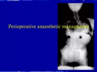
The. Scoliosis. Dr.Mouayad Kazem. Damascus Hospital. 27/4/2006. What is the Scoliosis ?. Lateral deviation of the vertebral line of the spine (measured > 10 °) Abnormal movement in 3 planes : Intervertebral extension Lateral intervertebral tilting Rotary component.

E N D
The Scoliosis Dr.Mouayad Kazem Damascus Hospital 27/4/2006
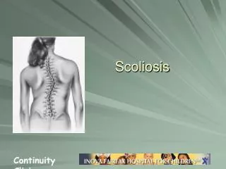
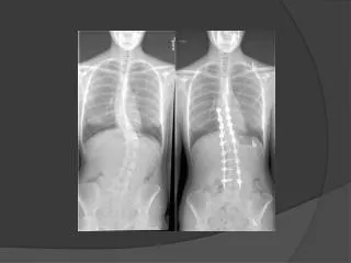
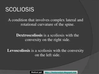
What is the Scoliosis ? • Lateral deviation of the vertebral line of the spine (measured > 10°) • Abnormal movement in 3 planes : • Intervertebral extension • Lateral intervertebral tilting • Rotary component
Classification • Idiopathic : • 80% • Any age – peaks in (1year _ 5-6 years _ 11 year) • Secondary : • Congenital • Neuromuscular • Hysterical
Idiopathic Scoliosis • Infantile Idiopathic Scoliosis • Birth – 3 years old • Juvenile Idiopathic Scoliosis • 4 – 10 years old • Adolescent Idiopathic Scoliosis • 11 year – skeletal maturity
Terms describing different types of Scoliotic curves • Structural curve • Nonstructural curve • Primary curve • Compensatory curve • lordoscoliosis • Kyphoscoliosis
Terms describing different types of Scoliotic curves • Cervicothoracic curve • Apex at C7 – T1 • Thoracic curve • Apex at T2 – T11 • Thoracolumbar curve • Apex at T12 – L1 or T12-L1 interspace • Lumbar curve • Apex at L1 – L4 • Lumbosacral curve • Apex at L5 or below
Terms describing different types of Scoliotic curves • Double curves • Double major curves • Double thoracic curves
Infantile Idiopathic Scoliosis • Birth – 3 years (most in first 6 months) • Left curve 90% • ♂>♀ ( 3 : 2 ) • 90% = self limited • Double major curves → severe deformity • Right thoracic curves + ♀→ bad prognosis.
Infantile Idiopathic Scoliosis • Clinical examination : • The spine , neurologic , associated deformities • Xray evaluation : • Cobb angle • RVAD • Phase of the rib head
Infantile Idiopathic Scoliosis • Rib Vertebral Angle Differencs ( RVAD) RVAD = RVA Concave – RVA Convex RVAD > 20° = progressive RVAD < 20° = self limited
Infantile Idiopathic Scoliosis • Phase of the rib head
Juvenile Idiopathic scoliosis • 12-21% • ♀< ♂(2:1→4:1) • Average age of Diagnosis : • ♀= 7 years • ♂ = 5 years • 70% progress → require treatment • Curve type resemble AIS : Right thoracic – DM
Juvenile Idiopathic scoliosis • Mehta classification : • Late-resolving idiopathic scoliosis • Benign progressive infantile idiopathic scoliosis • syndromic scoliosis • Syringomelic scoliosis • Early detected AIS
Juvenile Idiopathic scoliosis • Clinical examination : • Spinal deformities • Midline dimples • Hairy patches • Neurologic signs • Loss of abdominal reflexes • Absent gag reflex
Juvenile Idiopathic scoliosis • Investigations : • MRI : all of the patients <normal neurologics> Or : in patients <10 year + curve > 20° • Xray : • Cobb angle • RVAD : no benefit in prognosis But : used in predicting response to brace treatment
♂ + Left curve + cobb> 45° + kyphosis < 20° ↓ Progressive Juvenile Idiopathic scoliosis
Adolescent Idiopathic Scoliosis - AIS • ♀>♂(curve>45°= 9:1) • Discovered in school screening tests • No complaints ( cases of pain ) • AIS is a diagnosis of clinical and radiographic exclusion . • Left thoracic curves → syringomyelia
AIS – Before Skeletal Maturity • Factors predict progression : • Sex : Females • Remaining growth Assessment by : • Menarchal status ♀ • Risser sign • Tanner index • Peak Height Velocity PHV
AIS – Before Skeletal Maturity • Risser sign
AIS – Before Skeletal Maturity Tanner stages - Girls
AIS – Before Skeletal Maturity Tanner stages - Boys
AIS – Before Skeletal Maturity • Factors predict progression (cont.): • Curve size : • Risser 0 + premenarch + curve > 20° → high risk of progression • Curve pattern : • DM – Thoracic curves = high risk • Thoracolumbar • Lumbar = least risk
AIS – After Skeletal Maturity • Curves < 30° = unlikely to progress • Curves > 50° = worsen Thoracic curves = 1° / year • Mortality rate is the same • Infantile-Juvenile idiopathic scoliosis = ↑mortality Respiratory failure – cardiovascular disease • Chronic back pain • Lumbar osteoarthritis
Scoliosis screening • signs • Shoulder asymmetry • Unequal scapular prominence • Elevated or prominent hip • Greater space arm-body on one side • Head not centered over the pelvis • Adam forward bending test + • Scoliometer Recommendation for orthopedic referral = 5° trunk rotation
AIS - Etiology • Neurologic dysfunction • Vestibular-eye-proprioceptive systems • ↓ response to vibratory stimuli • Melatonin deficiency • Connective tissue abnormalities: • Difference in collagen • Ligamentum flavum • Paravertebral musculature • ↑ platelet calmodulin level • Genetic factors
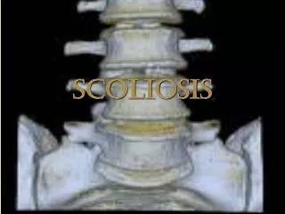
AIS - Pathophysiology • Changes are greatest at apex and diminish toward ends • In structural scoliosis : rotation of the vertebral body is to the convexity and the spinous process is to the concavity . • The scoliotic portion of the spine is lordotic in the sagittal plane
AIS - Patient evaluation • Complaint : • Body deformity • Pain : age>15 - risser≥ 2 – postmenarch – history of injury • Most common causes are : • Spondylolysis • Spondylolisthesis • Scheuermann’s kyphosis • Less common causes are: • Spinal cord syrinx • Disk herniation • Tethered spinal cord • Tumor • Respiratory symptoms • Neurologic deficit
AIS - Physical examination • Inspection : • Skin : midline hemangiomas – hair tufts – lumbar dimpling • Body asymmetry • Palpation : • Absence of a spinous process (spina bifida occulta) • Adam’s forward-bending test • Spinal balance
AIS - Physical examination Trunk balance Plumb line
AIS - Physical examination • Neurologic examination : • Patients reflexes • Superficial abdominal reflexes • Muscle test : • Range of motion • Hands and feet • Callus – nail bed irregularities.
AIS - Image study • Plain Xray : • Posteroanterior view (less radiation exposure) • Curve type and size • Trunk and vertebral balance • Skeletal maturity • Pelvic tilt • Appropriate fusion levels • Lateral view : • Lordoscoliosis • Hyper – hypo kyphosis • Spondylolisis - Spondylolisthesis
AIS - Image study • Plain Xray cont. • Bending view : • Curve flexibility • Surgical-nonsurgical indication • Stagnara view Stagnara view
AIS - Image study Curve size: Cobb angle
AIS - Image study Vertebral rotation
Sagittal balance on plain radiograph AIS - Image study
AIS - Image study • Dorsal kyphosis : • Apex at T4-T7 • Size 20-45° • Lumbar lordosis: • Apex at L3-L4 • Size 50-65° • 20% at L4-L5 • 40% at L5-S1 • Lumbar disks = 47° • vertebral bodies = 12° Balanced SVA = lumbar lordosis > thoracic kyphosis 20-30°
AIS - Curve types Ponseti – Friedman classification • Single major lumbar curve • Single major thoracolumbar curve • Combined thoracic and lumbar curves • Single major thoracic curve • Single major high thoracic curve • Double major thoracic curve
AIS – Lenk classification It’s a trial system combining curve type , the lumbar spine modifier and thoracic sagittal modifier eg. 1AN There are 42 different classifications
Scoliosis - Treatment Infantile idiopathic scoliosis: • Cobb <25° + RVAD <20 °→ observation • 5-10° increment of cobb/RVAD → treatment • Methods: • Serial casting • Milwaukee brace
Scoliosis - Treatment Juvenile idiopathic scoliosis • Curve <20° → observation • Curve >25° + flexible → Milwaukee brace • curve <35° + RVAD <20° = excellent prognosis • Curve >45° + RVAD >20° = poor prognosis • Curve >40-50° → Surgical treatment • Depends on : • Age • Spinal growth remaining < essential > • Expected loss of spinal growth • The aim is to halt progression of scoliotic curve. • Crankshaft phenomenon
Treatment – Juvenaile IS Surgical methods : • Instrumentation without fusion or with limited fusion • Hooks at the end vertebrae with rod • The rod is lengthened every 6-12 months • The same with localised fusion at the apex • Lessens curve progression • Luque rod and wires without posterior fusion • No need to lengthen the instrument • No need for external fixation • The same with anterior fusion at the apex