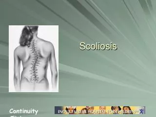
Scoliosis
370 likes | 1.81k Views
Scoliosis. Objectives. Know the etiology and natural history of scoliosis Describe how and when to examine for scoliosis Know how to determine the magnitude and pattern of spinal curvature Be familiar with treatment options. Epidemiology.

Scoliosis
E N D
Presentation Transcript
Objectives • Know the etiology and natural history of scoliosis • Describe how and when to examine for scoliosis • Know how to determine the magnitude and pattern of spinal curvature • Be familiar with treatment options
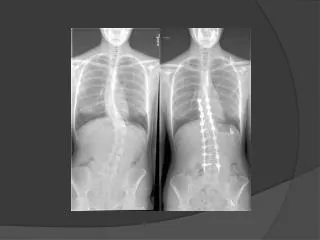
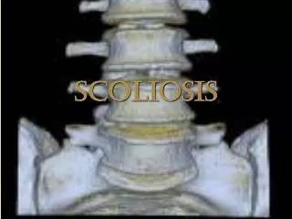
Epidemiology • Scoliosis is a lateral curvature of the spine greater than 10 degrees. • Idiopathic vs. Secondary • Idiopathic is the most common type. • Secondary causes include connective tissue, neurologic, and musculoskeletal disorders.
Classification • Idiopathic Scoliosis - defined by the age of onset • Infantile - birth to 3 years • Juvenile - 3 to puberty • Adolescent - after puberty • Adolescent Idiopathic Scoliosis is the most common type.
Etiology: AIS • No direct cause has yet been isolated. • Leading theory: Multigene dominant condition with variable phenotypic expression • Studies of twins have shown greater risk in monozygotic than dizygotic, and the rate of curve progression was nearly identical
Prevalence: AIS • Scoliosis is present in 2 to 4% of children between 10 and 16 years of age. • Girls tend to have more severe curves. • F:M ratio 1:1 in those with small curves (10 degrees) • F:M ratio increases to 10:1 in those with curves greater than 30 degrees
Diagnosis • Need to exclude secondary causes. • History: • family history • presence of pain and neurologic changes • bowel and bladder dysfunction
Physical Exam • Complete neurologic exam • Tanner staging - curve progression occurs most rapidly during stage 2 or 3 • Adam’s forward bend test
Adam’s Bend Test • Pt bends forward, spine horizontal to the floor, while holding palms together, arms extended. • Examine from side and behind the patient. • Look for a “rib hump” • Rib hump is a hallmark of a scoliotic curve greater than 10 degrees. Make sure pelvis is not dipping on one side AND leg length is symmetric
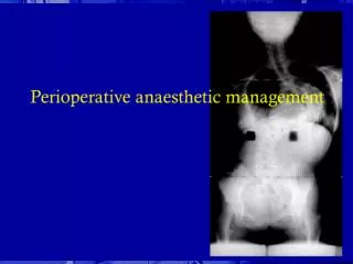
Imaging • Imaging is ordered for any patient with a lateral thoracic or lumbar spine curvature > 10 degrees. It should be considered in all patients with cervical curvature! • A single standing PA plain film of the spine is needed. • The degree of the curve is measured by the Cobb method. • 90% of curves are to the right!
Red Flags need MRI • A thoracic curve to the left • painful scoliosis • abnormal neurologic findings • untoward stiffness • deviation to one side during the bend test • sudden rapid progression in previously stable curve
The Cobb method • Choose the most tilted vertabrae above and below the apex of the curve. • Draw a line perpendicular to that vertabrae. • The angle created between these intersecting lines is the Cobb angle.
When do you observe vs. treat or refer? • What is the likelihood the curve will progress? • What degree of curvature leads to medical complications?
Will the curve progress? • Three factors involved in progression • patient’s gender • future growth potential • curve magnitude at time of diagnosis • Females are 10 times more likely to have progression than males. • The greater the growth potential and larger the curve = more likely to progress
How to determine growth potential? • Tanner staging - pt’s in stage 2 and 3 more likely to progress • Risser grade • based on ossification of iliac apophysis • graded from 0 (no ossification) to 5 (complete bony fusion)
The magic # is... 30 • Data from multiple studies has yielded the Risk of Curve Progression table. • The table assists in predicting progression and hence guiding treatment. • What is the risk for an 11 yo girl with a 25 degree curve and Risser grade 1?
Curve Progression • Curves 30 to 50 degrees progress an average of 10 to 15 degrees over a lifetime. • Curves > 50 at maturity progress steadily at a rate of 1 degree per year. • Curves less than 30 at bone maturity are unlikely to progress.
Medical Complications • At 100 degrees or greater: increased potential for life threatening effects on pulmonary function • Psychologic illness: seen in up to 19% of females with curves great than 40 degrees as adults.
Does Screening help? • AAOS recommends screening girls at ages 11 and 13; boys once at 13 or 14. • AAP recommends at 10, 12, 14, and 16. • But in fact... in 1996 the US Preventative Task Force found insufficient evidence for or against screening in asymptomatic pts. This was updated again in June 2004 with the same conclusion.
Treatment • Orthotic braces - 74% success rate at halting progression • Must be worn 20 hours a day, but most pts are not compliant. • Braces do not correct scoliosis. • Surgical therapy is definitive, but indicated only for those at 40 degrees or above
Conclusion • Adolescent Idiopathic Scoliosis is the most common type. • Overall, females more prone and tend to have more severe curves (to the right!). • Screening is of limited value. • There are extensive research based guidelines for predicting curve progression and treatment