Download

1 / 51
510 likes | 722 Views
The Elements of Wound Care. Brian K Wagner, DPM, CWS February 25, 2008. Objectives. Understand the wound evaluation process. Learn about the Core Healing Principles. Understand the wound healing process. Review questions from the certification process.

E N D
The Elements of Wound Care Brian K Wagner, DPM, CWS February 25, 2008
Objectives • Understand the wound evaluation process. • Learn about the Core Healing Principles. • Understand the wound healing process. • Review questions from the certification process. • Learn about growth factors involved in wound healing. • Learn about the benefits of Dermagraft on wound healing. • Discuss select articles.
Patient Factors • General appearance • Mental status • Body type • Age • Previous amputation • Ethnicity • Mobility • PMH
Patient Factors • Social History • Occupation • Smoking • ETOH • IVDA • Pain • Past surgical history
Patient Factors • Wound History • Timing • Location • Previous ulcer history • Past treatments • Size appearance • Complications/infections • Drainage • Pain
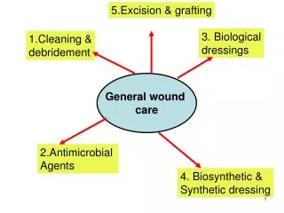
Wound Factors • Periwound Skin • Nonblanchableerythema • Underminging • Pain • Dry, cracked, fissured • Edematous • Rolled-over-edges
Wound Factors • Wound bed assessment • Moisture balanced • Presence of necrotic tissue • Granulation tissue • Consider biopsy • Abnormal appearance • Wound present >6 months
Wound Factors • Wound bed analysis • Depth • Length • Width • Location • Duration • Exudation • Quality of wound bed tissue
Macrolevel • Macro and Microcirculation • Palpable pulses • Non-invasive examinations • Vascular consultation • Bacterial bioburden • Quantitative biopsy – gold standard
Macrolevel • Pressure reduction • Entire wound • Dressing may cause increase in pressure and local ischemia • Nutritional assessment • Pre-albumin • Total lymphocyte count • Albumin level • Immunosuppression • Steroids, chronic diseases, HIV
Albumin vs. Prealbumin • Both are indicators of visceral protein status. • Determine the degree of protein malnutrition. • Both are a negative acute-phase reactant • Meaning levels could reflect inadequate nutrition or inflammatory stress.
Albumin • Maintains colloidal osmotic pressure. • Depleted albumin levels may cause edema, ascites, or pulmonary edema. • A late indicator of malnutrition (half life is 20 days) • Levels below 3.0gm/dL : • is associated with tissue edema • Increased risk of infection, morbidity, and mortality. • Impairs or prevents wound healing and decreases wound tensile strength.
Prealbumin (Transthyretin) • Shorter half-life of 2 days • More sensitive measure of nutritional status. • Mild depletion is <15mg/dL • Severe depletion is <5mg/dL • Chronic renal failure may falsely elevate levels.
Microenvironment • Microcirculation • TcpO2 • Intrvitalcapillaroscopy • Noninvasive using a microscope • Laser doppler
Laser Doppler • Low-intensity laser light (helium-neon) • Measures backscattering created by moving RBC’s • Two parameters • Number of shifted photons • Relates to the concentration of moving RBC • Mean doppler shift of the photons • Relates to the blood cell velocity
Microenvironment • Excessive inflammatory mediators • Bacterial byproducts cause disturbances in the normal healing process and may create a chronic wound. • Proliferative capacity of the host to heal. • Growth factor deficiencies • Excessive MMPs
Normal Healing Process • Hemostasis • Inflammation • Proliferation • Epithelialization • Remodeling
Hemostasis • Injury -> bleeding -> release of clotting factors -> platelet aggregation -> fibrin clot formation • Clot stops blood loss and acts as a temporary bacterial barrier
Hemostasis • Growth factors are released from α-granules of platelets which initiate the entire wound healing cascade. • Hemostasis initiates the wound healing process.
Inflammation • Initial vasodilation occurs after the vasoconstriction of the hemostasis phase. • Mast cells release: prostaglandins, leukotrienes and histamine. • Neutrophils and monocytes migrate into the wound caused by chemoattractants. • Monocytes become phagocytic and are called macrophages.
PMNs • First cells to arrive • Provide an initial barrier to bacterial invasion. • Phagocytize bacteria and debris in the wound.
Inflammatory • Growth factors released by macrophages cause: • Attraction of fibroblasts • Attraction of epithelial cells • Attraction of vascular endothelial cells • Macrophages regulate the entire wound healing process.
Inflammatory • Controls bleeding • Creates a clean wound bed • Lasts for 3 days • If necrosis or infection are present the inflammatory phase is prolonged and wound healing is delayed.
Proliferative • Fibroplasia – reinforcement of injured tissue • Neovascularization • Reepithelialization
Proliferative • Fibroblasts • synthesize collagen and other connective tissue • Respond to cytokines, PDGF, activated neutrophils and activated macrophages and migrate into the wound.
Proliferative • Wound contraction (3 theories) • Myofibroblasts – contain α-smooth muscle actin capable of generating a strong contraction on the wound. • Fibroblasts act in concert to reorganize the wound causing it to shrink. • New collagen fibers have cellular forces which produces a pulling force on the wound.
Proliferative • Epithelialization • Occurs over moist, vascular wound beds. • Full-thickness wounds heal from the periphery inward. • Rolled edges prevent migration of epithelial cells and must be excised or debrided.
Remodeling • Collagen synthesis and breakdown. • Lasts up to 1 year. • Tensile strength is 80% of nonwounded tissue
Acute vs. Chronic • Chronic wounds • have no clot formation or breakdown. • Typically occur in compromised hosts. • Vicious cycle is created: inflammatory cells release proinflammatory cytokines -> breakdown into chemoattractants -> attract additional inflammatory mediators -> causing further tissue breakdown.
Acute vs. Chronic Wounds • Acute • High mitotic activity • Therapeutic levels of inflammatory cytokines • Low levels of proteases • Chronic • Low mitotic activity • Increased levels of inflammatory cytokines • High levels of proteases
Questions • Which of the following cells appear to be the principal mediators for full-thickness wound repair? A. Endothelial cells and macrophages • Neutrophils and platelets • Fibroblasts and neutrophils • Platelets and macrophages
Questions • Contraction plays an important role in healing for? • Wounds healing by primary intention. • Wounds healing by secondary intention. • Superficial abrasions • Partial-thickness wounds
Question • A significant source of growth factors is the? • Endothelial cell • Fibroblast • Monocyte • Macrophage
Questions • The molecular environment of a chronic wound is characterized by? • Tight regulations of levels of inflammatory cytokines • Increased levels of proteases • Increased levels of protease inhibitors • Excessive collagen synthesis
Questions • Human dermis is mostly composed of? • Type 1 collagen • Type 2 collagen • Type 3 collagen • Type 4 collagen
Questions • What initiates the wound healing process? • Bacteria • Wound drainage • Debridement • Proper dressing selection • Hemostasis
Questions • Epidermal migration needs? • Necrotic wound with drainage • Dry eschar • Moist wound • Good blood supply • Both c and d.
Questions • How does one calculate total lymphocyte count (TLC)? • Why is debridement so important?
Wound Healing Actions • Chemotactic migration -> replacement of epidermal and dermal cells -> growth of new blood vessels -> formation of scar tissue -> remodeling of scar tissue
Hemostasis • Platelet derived growth factor (PDGF) • Transforming growth factor-beta (TGF-β) • Epidermal growth factor (EGF) • Insulin-like growth factor-I (IGF-I)