Download
1 / 1
10 likes | 273 Views
Transmissible Spongiform Encephalopathy Prion Protein Diseases Lisa Kennedy, Dylan Bradford, Madi Hoagland Henefield, Anders Ohman Advisor: Dr. Todd Livdahl. Molecular Biology of the Pathogenesis of TSE. Background. Transmission Routes.

E N D
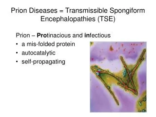
Transmissible Spongiform Encephalopathy Prion Protein Diseases Lisa Kennedy, Dylan Bradford, Madi Hoagland Henefield, Anders Ohman Advisor: Dr. Todd Livdahl Molecular Biology of the Pathogenesis of TSE Background Transmission Routes Prion diseases (formally, Transmissible Spongiform Encephalopathies) are neurodegenerative conditions in mammals that are hypothesized to be caused by an infectious protein agent without the involvement of nucleic information or additional transport vectors. The theorized prion agent is a modified form of the cell membrane protein PrPC, which is misfolded into the pathogenic form of the protein, which is referred to as PrPSc. These prion proteins are able to convert normal protein molecules into the prion form, and the proteins are extremely resistant to denaturation by temperature or chemical agents, making them both difficult to destroy once in a host as well as capable of surviving in an exposed environment for an extended period of time. Prion diseases have been documented for decades (centuries, in the case of Scrapie), but the discovery of the prion protein itself was made as recently as 1997 by Stanley B. Prusiner, who won a Nobel Prize for his work. The mechanics of the disease are still largely unknown. They can affect many lineages of mammals, from rodents to ruminants, as well as humans. Prion disease in livestock has serious implications for human food source populations, as well as for potential infection in humans, as the Bovine Spongiform Encephalopathy may potentially be linked to a variant of Creutzfeldt-Jakob Disease in humans. There is no current cure, inoculation, or treatment for prion diseases, and livestock protection strategies largely include quarantine and destruction of infected organisms. Symptoms: TSEs cause the formation of holes in the host nervous tissue, causing a sponge-like appearance from which the term ‘spongiform’ is derived. Damage to the nervous tissue causes a number of deteriorative cognitive and behavioral conditions in prion hosts of all susceptible species, typically including dementia, acute mood changes, and issues with physical coordination and control. It is commonly held that prion diseases are mainly transmitted between animals via ingestion or orally. A number of ingestion routes have been identified that cause disease in humans as well as other animals including deer, elk, and cattle. The most common and surest way is to ingest tissue from an infected animal, the brain tissue having the highest concentration of dangerous prions. Omnivorous and carnivorous animals are susceptible to this mode of infection, and exhibits a very real danger to human hunters of deer and elk. A study of humans that were exposed to Chronic Wasting Disease (a closely related prion disease) via hunted deer at a wild game feast suggested two levels of risk, exposure and ingestion (Garruto et al. 2008). Those subject that had actually ingested venison that could have come from an infected deer were said to be of high-risk and are currently under close watch in case CWD is found to transmit between deer and humans. For herbivores it was found that prions, in the form of Scrapie Agent 263k, are able to stay dormant in soil for at least as long as 29 months and cause infection (Seidel et al. 2007). The study was done using Syrian hamsters and PrPSC were found to infect and proliferate within the hamsters when fed soil samples. Aqueous extract was also shown to induce disease in the reporter animals. These findings suggest a highly dangerous scenario for cattle and sheep flocks. Prions can be deposited by decaying animals, as well as infected organs including placenta, amniotic fluid, and possibly urine (Gregori et al. 2008), making grazing animals highly susceptible. There still remains much research to be done on prion diseases as a whole including transmission routes. The way they are passed between animals is very important to determine if only to protect humans from possible cross-species infection. Given the amount of red meat consumed in the United States, a large portion of infected but undetected meat due to a lack of knowledge could cause a large loss of life. Prion disease are unique in that they are transmissible particles that lack any form of nucleic acid, and yet are still infectious (Prusiner, 1998). While prion diseases are not completely understood, it is clear the disease is caused by an accumulation in the brain of a misfolded cellular protein known as the prion protein (PrPc) (Campana et al., 2008). The normally occurring prion protein is converted into the modified infectious protein PrPSc posttranslationally. During this process PrPc misfolds into PrPSc which contains a high number of β-sheets. The process during which the normal protein changes to the abnormal takes place when some of the α-helices and coils of its structure refold into β-sheets. This structural alteration causes less understood but still profound changes in the chemical properties of the PrP. The changes that result in the PrPSc have been shown to act as a template upon which PrPc is refolded into PrPSc (Prusiner, 1998). This is how the misfolded protein is able to become infectious to other prion proteins around it. Furthermore, PrPSc has shown the ability to resist degradation even after the death of its host for up to months or even years at a time, and still be infectious to a new host if picked up. Modeling the System The model below calculates the number of new cases of BSE that result from the use of infected BSE cattle in cattle feed. The practice of “recycling” cattle was relatively common before the outbreaks of BSE in the UK. This STELLA model (fig. 1) utilizes data used in a published model used to calculate the hypothetical reproductive ratio of BSE, as well as actual data on the cattle population of the UK in 1986. Types of TSE Prion disease occurs in a number of different mammals, and strains of prion disease typically only affect peers in the species and closely-related lineages (Prusiner, S.B. et al 1991). There are three primary categories of prion diseases: Sporadic, where the cause is unknown ; Familial, where the disease is caused by the genetics of the host; and Acquired, which is transmissible between organisms, either directly or through the environment. There are four primary prion diseases that affect humans: Creutzfeldt-Jakob Disease, Kuru, Fatal Familial Insomnia, and Gerstmann-Straussler-Scheinker Syndrome. CJD has variant strains in all three categories, while Kuru is an Acquired disease and both FFI and GSSS are Familial (Soldevila, M. et al 2006). Symptoms of CJD, the most common human prion disease with 1-2 instances in a million people annually, has the symptoms of dementia, issues with coordination, and visual hallucinations. GSSS shares the symptoms of dementia and coordination problems, but also results in difficulty speaking. Sufferers of Kuru have laughing fits and trembling fits. FFI causes insomnia, hallucinations, and also dementia (Monari, L. et al 1994). Deer, Elks, and Moose are affected by Chronic Wasting Disease, an Acquired condition that is transmissible directly or through contact with a shared environment (Joly, D.O. et al 2003). CWD was first contained to the Central United States, but has since spread to multiple areas of the US and Canada, including game parks. It causes changes in host mood, diet, and behavior. Sheep and Goats are affected by Scrapie, the oldest-known prion disease first named in 1732 (www.defra.gov.uk). Scrapie affects both the nervous system as well as the tonsils and the distal ileum, and causes behavioral abnormalities such as the compulsive scraping of the host on objects in the environment from which the diseases’ name is derived. Although found in Europe and in America, successful quarantine has kept the disease out of Oceanian populations (Parsonson, I.M. 1996). Cows are affected by Bovine Spongiform Encephalopathy. While BSE (a.k.a. Mad Cow Disease) is a strictly Familial disease, domestic animal practices for feeding livestock resulted in direct consumption of infected brain tissue as animal feed, and therefore spread the disease from individual to individual (Prusiner, S.B. 1997). BSE leads to the formation of protein fibers in the brain, leading to the presence of holes in the tissue. Symptoms include nervous or aggressive behavior, weight loss, and coordination problems, but can take years to actually appear. 372 1947: Transmissible mink encephalopathy 1967: Chronic Wasting Disease CWD: captivity 1921: Creutzfeldt-Jakob disease (CJD) 1996: Variant Creutzfeldt-Jakob Disease 1981: CWD wild populations TIMELINE Kuru: 1959 1732*: Scrapie 1936: Gerstmann Straussler-Scheinker Syndrome (GSSS) 1974: Fatal Familial Insomnia 1986: BSE 1997: Prion protein discovered*** 2003: Feline spongiform encephalopathy *Timeline not to scale **The dates indicate descriptions and identifications of the respective diseases. At the earlier dates, cause of disease by the prion protein was not known ***By Stanley B. Prusiner winner of the Nobel Prize in Physiology or Medicine 1997