Arrythmia Interpretation (cont’d)
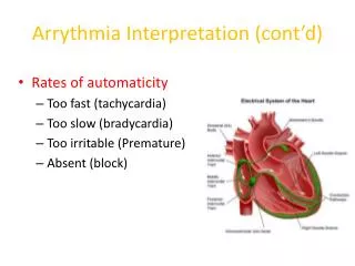
Arrythmia Interpretation (cont’d). Rates of automaticity Too fast (tachycardia) Too slow ( bradycardia ) Too irritable (Premature) Absent (block). Interpreting Arrhythmias. 1. Calculate the heart rate 2. Assess the rhythm 3. Identify the P waves 4. Assess QRS shape and duration

Arrythmia Interpretation (cont’d)
E N D
Presentation Transcript
Arrythmia Interpretation (cont’d) • Rates of automaticity • Too fast (tachycardia) • Too slow (bradycardia) • Too irritable (Premature) • Absent (block)
Interpreting Arrhythmias 1. Calculate the heart rate 2. Assess the rhythm 3. Identify the P waves 4. Assess QRS shape and duration 5. Assess relationship between P waves and QRS complexes 6. Name the arrhythmia
Normal Sinus Rhythm • Normal ECG tracing depicting a normal rhythm of electrical conductivity through the heart
(Respiratory) Sinus Arrhythmia • All criteria of normal rhythm except heart and pulse rates increase with inspiration and decrease with expiration • Normal finding in brachycephalic breeds and in chronic respiratory disease • Increased number of cardiac cycles during inspiration; decreased number during expiration • Originates in the SA node
What is the normal HR for dogs and cats? • Dogs: 70 – 160 BPM • Cats: 150 – 210 BPM
Sinus Bradycardia • Regular sinus rhythm but heart rate is below normal • Dogs under 45 lb: HR less than 70 bpm • Dogs >45 lb: HR < 60 BPM • Cats: 100 BPM or less • CS: weakness, hypotension, syncope
Sinus Tachycardia • Regular sinus rhythm with increased ventricular rate • Dogs less than 45 lb; HR >180 BPM • Dogs more than 45 lb; HR >160 BPM • Cats: HR greater than 240 BPM • Causes include: pain, fever, anemia, excitement, hyperthyroidism
Atrial Premature Complexes • Premature atrial impulses originating from ectopic atrial site other than SA node • Seen in dogs and cats with atrial enlargement, electrolyte disturbances, drug reactions, congenital heart disease, and neoplasia; a normal variation in older animals • Premature P wave • QRS complexes are normal unless the P wave is so immature that it overlaps to varying degrees
Atrial Premature contraction/complexes Represent premature P wave/s
Atrial Tachycardia • Rapid regular rhythm originating from an atrial site other than the sinus node • May be seen in dogs with severe heart disease and in cats with cardiomyopathy or hyperthyroidism
Atrial Flutter • Appears as a regular, sawtooth formation between the QRS complexes. • Occurs when the ventricular rate differs from the atrial rate. • Atrial flutter is the precursor to atrial fibrillation.
Fibrillation is the rapid, irregular, and unsynchronized contraction of muscle fibers.
Atrial Fibrillation • Caused by numerous disorganized atrial impulses frequently bombarding the AV node. • Ventricular depolarization rate is irregular and rapid. • No P waves are evident; replaced by numerous f waves. • QRS complexes may be normal or wide and of varying amplitude
Premature Ventricular Complexes (PVCs) • “Premature beats” - Cardiac impulses initiated within the ventricles instead of the sinus node • Ventricle discharges before the arrival of the next anticipated impulse from the SA node. • Can occur at any rate but pose a greater danger with tachycardia. • Associated with congenital defects, cardiomyopathy, GDV, drug reactions, cardiac neoplasia, anemia, acidosis, hyperthyroidism, hypokalemia
PVCs (cont’d) • The P wave is often not seen on the ECG tracing. • A wide, distorted QRS complex is also evident • The beat preceding the PVC and the beat following are usually equal to the time of two normal beats.
Ventricular Tachycardia • A series of four or more PVCs in a row . • Potentially life threatening.
Ventricular Fibrillation • The mechanical pumping of the heart is not evident on the ECG. • The ECG has bizarre baseline with prominent undulations due to weak and uncoordinated ventricular contractions. • Low to absent cardiac output. • Associated with shock, trauma, electrolyte imbalances, drug reactions, electric shock, hypothermia, cardiac surgery. • Rapidly fatal
Ventricular Fibrillation • There are no recognizable P or QRS complexes. • Irregular, chaotic, deformed reflections of varying width, amplitude, and shape. • Unless controlled immediately, ventricular fibrillation will result in cardiac arrest.
Sinus Arrest or Block • Normal sinus rhythm interrupted by an occasional prolonged failure of the SA node to initiate an impulse. • Conduction disturbance in which normal sinus rhythm is interrupted by an occasional, prolonged failure of the impulse generated by the SA node to reach the atria.
Heart Block • Electrical impulse is not transmitted through the heart.
First Degree AV Block • Delay in conduction of an impulse through the atrioventricular junction and Bundle of His. • The PR interval is longer than normal. • This type of heart block is a result of a minor conduction defect. • Seen in older patients secondary to degenerative changes in the conduction system.
Second Degree AV Block • Some atrial pulses are not conducted through the AV node and therefore do not cause depolarization of the ventricles. • There are two types: • Type I (Mobitz type I or “Wenckebach” AV block): progressive lengthening of the PR interval on successive beats and then P waves occurring without QRS complexes. • P waves occurring without QRS complexes are called “dropped beats”
Second Degree AV Block (cont’d) • Type II: A constant PR interval that is usually of normal duration with random dropped beats.. • In the case of type 2 block, atrial contractions are not regularly followed by ventricular contraction
Third degree AV block • The cardiac impulse is completely blocked in the region of the AV junction and/or all bundle branches. • Also known as a complete heart block; the most severe heart block. • No relationship between P waves and QRS complexes; atria and ventricles each beat independently. • Atrial rate is normal.
Asystole (Flat line) Cardiac Arrest: No cardiac electrical activity, no cardiac output or blood flow. At this point the heart will not respond to defibrillation. Causes: hypoxia, hypothermia, hypoglycemia, or an electrode has fallen off (hopefully)
Asystole (Flat line) Medications of choice: Epinephrine or Atropine along with manual chest compressions.